. 2016 Mar;20(3):500-9.
doi: 10.1007/s11605-015-3062-2. Epub 2015 Dec 29.

Bile Salts at Low pH Cause Dilation of Intercellular Spaces in In Vitro Stratified Primary Esophageal Cells, Possibly by Modulating Wnt Signaling

Affiliations

Bile Salts at Low pH Cause Dilation of Intercellular Spaces in In Vitro Stratified Primary Esophageal Cells, Possibly by Modulating Wnt Signaling

Sayak Ghatak et al. J Gastrointest Surg. 2016 Mar.

Abstract

Background: The presence of dilated intercellular spaces in the stratified squamous lining of the esophagus is the pathognomonic feature of reflux esophagitis secondary to gastroesophageal reflux disease (GERD). In addition to stomach acid, bile salts are major constituents of gastroesophageal refluxate. The aim of our study was to determine the effect of bile salts cocktail at different pHs on epithelial junctions in an in vitro transwell model of stratified esophageal squamous epithelium.

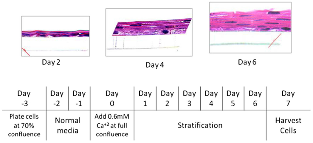
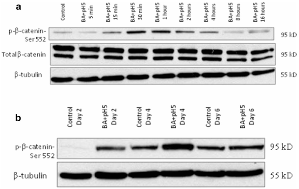
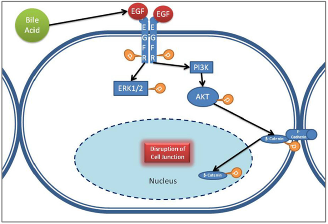
Discussion: Human telomerase reverse transcriptase (hTERT) immortalized primary esophageal EPC1 cells were grown on polyester transwell surfaces in calcium-enriched media. The cells exhibited gradual stratification into an 11-layered squamous epithelium over 7 days, together with epithelial barrier function as indicated by increased transepithelial electrical resistance (TEER). This stratified epithelium demonstrated well-formed tight junctions, adherens junctions, and desmosomes as visualized by immunofluorescence and electron microscopy. When exposed to short pulses of bile salts at pH 5, but not either condition alone, there was loss of stratification and decrease in TEER, concomitant with disruption of adherens junctions, tight junctions, and desmosomes, leading to the appearance of dilated intercellular spaces. At the cellular level, bile salts at pH 5 activated the Wnt pathway (indicated by increased β-catenin Ser552 phosphorylation).

Conclusion: In conclusion, in our in vitro transwell model bile salts at pH 5, but not bile salts or media at pH 5 alone, modulate Wnt signaling, disrupt different junctional complexes, and cause increased permeability of stratified squamous esophageal epithelium. These changes approximate the appearance of dilated intercellular space similar to that found in GERD patients.

Keywords: Bile salt; Dilated intercellular spaces; Gastroesophageal reflux disease.


Figures

Fig. 1
Fig. 1
Dilated intercellular spaces in patient biopsies—biopsies taken from the lower esophagus of patients diagnosed with clinical GERD showed discernible dilatation of intercellular spaces with hematoxylin and eosin staining under both low and high magnification, whereas biopsies taken from non-GERD individuals showed tight intracellular connection with virtually no intercellular spaces (Fig. 1)
Fig. 2
Fig. 2
EPC1 cells stratify in presence of calcium on polyester transwells—hematoxylin and eosin staining of EPC1 cells on transwell gradually stratifies in 6 days of culture in media enriched with calcium. The culture protocol is shown in the schema below
Fig. 3
Fig. 3
Treatment with bile acid at pH 5 decreases epithelial resistance—transepithelial electrical resistance increased through day 1–6 as EPC1 cells stratified on transwells (a, bblue line). TEER reduced overnight after the third pulse treatment with cocktail of bile salts at pH 5 (a, byellow line), but with neither alone (ared and green lines)
Fig. 4
Fig. 4
Bile salts at pH 5 cause loss of stratification—electron microscopy of EPC1 cells grown on transwells after 7 days of treatment. Apical side is on the top, and basal side along with the polyester filter can be seen at the bottom. Nucleated basal cells can be seen at the bottom, and non-nucleated, flattened, terminally differentiated keratinocytes can be seen on the top. Cells treated with the control media, bile salts at pH 7.4, show similar levels of stratification. Cells treated with bile salts at pH 5 show considerable loss of stratification. pH 5 also shows some loss of stratification
Fig. 5
Fig. 5
Bile salts at pH 5 cause desmosomal disruption—disruption of desmosomes (solid arrow) as well as DIS (patterned arrow) is markedly visible at higher magnification after treatment with bile salts at pH 5 and at pH 7.4 to lesser extent. The desmosomal architecture remains intact after treatment with pH 5 media, similar to the control
Fig. 6
Fig. 6
Bile salts at pH 5 cause desmosomal disruption—in a, adherens junction protein E-cadherin is visualized in red, and tight junction protein zonula occludin-1 is visualized in green under confocal microscopy. Treatment of bile salts at pH 5 on stratified EPC1 cells on transwells caused disruption of cell-cell junctions, whereas pH 5 only or bile salts at neutral pH has no effect on the cell-cell junctions. In b, adherens junction proteins p120 catenin is visualized in red, and tight junction protein occludin is visualized in green under confocal microscopy. In c, adherens junction proteins AF6 is visualized in green, and adherens junction-associated proteins α-catenin and β-catenin are visualized in red under confocal microscopy. Both figures indicate treatment with bile salts at pH 5 on stratified EPC1 cells on transwells caused disruption of cell-cell junctions
Fig. 7
Fig. 7
Bile salts at low pH activate WNT pathway—bile salts at pH 5 caused β-catenin serine residue-552 phosphorylation in EPC1 cells on transwells, following 30 min of treatment, indicating WNT activation, with maximum phosphorylation seen at 1 h (a). Activation persisted with longterm treatment on days 2, 4, and 6 of treatment (b). Total β-catenin level remained unchanged. β-tubulin served as the loading control
Fig. 8
Fig. 8
Hypothetical sequence of events leading to junction disruption—at basal condition in a terminally differentiated stratified squamous cell, WNT signaling is inactive, and β-catenin binds to E-cadherin and is tethered to the adherens junction. Bile salts, possible by activating EGFR pathway and downstream Akt phosphorylation, might cause β-catenin phosphorylation at Ser552 residue triggering its release and subsequent disruption of the adherens junction complex

References

    1. El-Serag HB, Sweet S, Winchester CC, and Dent J, Update on the epidemiology of gastro-oesophageal reflux disease: a systematic review. Gut, 2013. - PMC - PubMed
    1. DeVault KR, Castell DO, and American College of G, Updated guidelines for the diagnosis and treatment of gastroesophageal reflux disease. Am J Gastroenterol, 2005. 100(1): p. 190–200. - PubMed
    1. Souza RF, Krishnan K, and Spechler SJ, Acid, bile, and CDX: the ABCs of making Barrett’s metaplasia. Am J Physiol Gastrointest Liver Physiol, 2008. 295(2): p. G211–8. - PubMed
    1. Kauer WK, Peters JH, DeMeester TR, Feussner H, Ireland AP, Stein HJ, and Siewert RJ, Composition and concentration of bile acid reflux into the esophagus of patients with gastroesophageal reflux disease. Surgery, 1997. 122(5): p. 874–81. - PubMed
    1. Orlando RC, Powell DW, and Carney CN, Pathophysiology of acute acid injury in rabbit esophageal epithelium. J Clin Invest, 198168(1): p. 286–93. - PMC - PubMed

LinkOut - more resources