. 2016 Mar;202(3):903-10.
doi: 10.1534/genetics.115.182469. Epub 2015 Dec 29.

Redundant Regulation of Cdk1 Tyrosine Dephosphorylation in Saccharomyces cerevisiae

Affiliations

Redundant Regulation of Cdk1 Tyrosine Dephosphorylation in Saccharomyces cerevisiae

Erin K Kennedy et al. Genetics. 2016 Mar.

Abstract

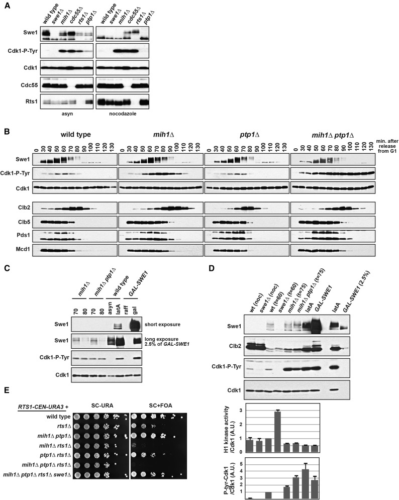
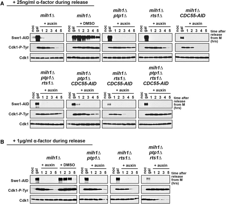
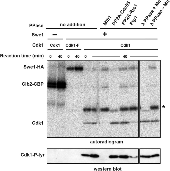
Cdk1 activity drives both mitotic entry and the metaphase-to-anaphase transition in all eukaryotes. The kinase Wee1 and the phosphatase Cdc25 regulate the mitotic activity of Cdk1 by the reversible phosphorylation of a conserved tyrosine residue. Mutation of cdc25 in Schizosaccharomyces pombe blocks Cdk1 dephosphorylation and causes cell cycle arrest. In contrast, deletion of MIH1, the cdc25 homolog in Saccharomyces cerevisiae, is viable. Although Cdk1-Y19 phosphorylation is elevated during mitosis in mih1∆ cells, Cdk1 is dephosphorylated as cells progress into G1, suggesting that additional phosphatases regulate Cdk1 dephosphorylation. Here we show that the phosphatase Ptp1 also regulates Cdk1 dephosphorylation in vivo and can directly dephosphorylate Cdk1 in vitro. Using a novel in vivo phosphatase assay, we also show that PP2A bound to Rts1, the budding yeast B56-regulatory subunit, regulates dephosphorylation of Cdk1 independently of a function regulating Swe1, Mih1, or Ptp1, suggesting that PP2A(Rts1) either directly dephosphorylates Cdk1-Y19 or regulates an unidentified phosphatase.

Keywords: Cdc25/Mih1; Cdk1; PP2A; Wee1/Swe1; mitosis.


Figures

Figure 1
Figure 1
PTP1 regulates Cdk1-Y19 phosphorylation in vivo. (A) mih1∆, cdc55∆, rts1∆, and ptp1∆ cells have increased Cdk1-Y19 phosphorylation. Wild-type, swe1∆, mih1∆, cdc55∆, rts1∆, and ptp1∆ cells were grown at 25° asynchronously (asyn) or arrested in mitosis with nocodazole, harvested, lysed, and blotted with the indicated antibodies. (B) Wild-type, mih1∆, ptp1∆, and mih1ptp1∆ cells were grown at 25°, arrested with mating pheromone, and released into the cell cycle at t = 0. Cells were harvested, lysed, and blotted with the indicated antibodies. (C) A comparison of Cdk1-Y19 phosphorylation in mih1∆ and mih1ptp1∆ cells in samples from B at t = 70 min and t = 80 min, wild-type cells treated with latA (2.5 µM), and cells overexpressing Swe1 from a GAL-SWE1 gene that replaces the endogenous SWE1. Cells arrested by latA or Swe1 overexpression have significantly more Swe1 than mih1∆ and mih1ptp1∆ cells and have slightly higher levels of phosphorylated Cdk1-Y19. (D) Differences in Cdk1-Y19 phosphorylation correlate with changes in Cdk1/Clb2 histone 1H-kinase activity. Wild-type, swe1∆, mih1∆, and mih1ptp1∆ cells were grown in nocodazole (noc) or arrested with mating pheromone (αf; 100 ng/ml) and released into the cell cycle at t = 0 and either harvested at the time of peak Cdk1-Y19 phosphorylation (t = 60 or 75 min post-release) or incubated with latA for 2 hr. GAL-SWE1 cells were grown in YEP + 2% raffinose, arrested in nocodazole, and then grown for 90 min after the addition of 2% galactose. Cells were harvested, lysed, and blotted with the indicated antibodies (top panels), or Cdk1/Clb2 complexes were immunoprecipitated with anti-Clb2 antibody and blotted with anti-Cdk1 and anti-Cdk1-P-tyr antibodies, or the Clb2-associated histone H1 kinase activity was assayed. The indicated ratios [average (±SEM) of three independent experiments] were calculated and normalized so that the ratio of synchronous mitotic wild-type cells (t = 60 min) is set to 1. (E) mih1rts1∆ and mih1ptp1rts1∆ cells are inviable, and this inviablity is rescued by swe1∆. Tenfold serial dilutions of the indicated strains were spotted on SC-URA or SC + FOA medium and grown at 25°. All strains harbor a RTS1-CEN-URA3 plasmid, and their dependence on RTS1 was examined by selecting for loss of the plasmid using 5-FOA, which counterselects for URA3 cells.
Figure 2
Figure 2
MIH1, PTP1, and RTS1 redundantly regulate Cdk1-Y19 dephosphorylation. The indicated strains were grown overnight in YEP + raffinose at 25° and arrested in mitosis with nocodazole (noc), and then galactose was added to induce high levels of Swe1-AID and Cdk1-Y19 phosphorylation. After 1 hr of induction (gal), cells were released from the nocodazole arrest, dextrose was added to repress transcription of GAL-SWE1-AID, and auxin was added to induce Tir1-dependent degradation of Swe1-AID and Cdc55-AID, if present in the strain. Control mih1∆ cells also were released into medium containing dextrose and vehicle (DMSO). ⍺-factor was added 90 min after release at (A) 25 ng/ml or (B) 1 µg/ml in order to rearrest the cells in the subsequent G1. Cells were harvested at the indicated times for immunoblotting with the indicated antibodies.
Figure 3
Figure 3
Mih1 and Ptp1 dephosphorylate Cdk1-Y19 in vitro. Purified Cdk1/Clb2-CBP was phosphorylated by bead-bound Swe1-HA in the presence of γ-[32P]ATP. Following phosphorylation, Cdk1/Clb2-CBP was removed from the beads and mixed with purified phosphatases and an excess of unlabeled ATP to prevent rephosphorylation of Cdk1 by γ-[32P]ATP and Swe1-HA that was washed off the beads. Reactions were run on a polyacrylamide gel and either dried and exposed to a phosphorimager screen or transferred to nitrocellulose and immunoblotted with anti-Cdk1-P-Y19 antibody. In the absence of Swe1-HA, Clb2 is heavily phosphorylated by Cdk1. In the presence of Swe1-HA, Clb2 is not phosphorylated, showing that Cdk1 is completely inhibited by Swe1-dependent phosphorylation of Y19. Swe1 does not phosphorylate Cdk1-F, but Cdk1-F phosphorylates Swe1 (Harvey et al. 2005). After incubation for 40 min at 25°, purified Mih1 and Ptp1 (see Figure S3) can dephosphorylate Cdk1-Y19 in vitro, but PP2ACdc55, PP2ARts1, and lambda phosphatase (λ-PPase) cannot. PP2ACdc55 and λ-PPase can effectively dephosphorylate a fortuitous phosphorylated background protein (indicated with an asterisk), providing a control indicating that both PP2ACdc55 and λ-PPase are active.

References

    1. Booher R. N., Deshaies R. J., Kirschner M. W., 1993. Properties of Saccharomyces cerevisiae wee1 and its differential regulation of p34CDC28 in response to G1 and G2 cyclins. EMBO J. 12: 3417–3426. - PMC - PubMed
    1. Cayla X., Goris J., Hermann J., Hendrix P., Ozon R., et al. , 1990. Isolation and characterization of a tyrosyl phosphatase activator from rabbit skeletal muscle and Xenopus laevis oocytes. Biochemistry 29: 658–667. - PubMed
    1. Costanzo M., Baryshnikova A., Bellay J., Kim Y., Spear E. D., et al. , 2010. The genetic landscape of a cell. Science 327: 425–431. - PMC - PubMed
    1. Deibler R. W., Kirschner M. W., 2010. Quantitative reconstitution of mitotic CDK1 activation in somatic cell extracts. Mol. Cell 37: 753–767. - PMC - PubMed
    1. Dunphy W. G., Kumagai A., 1991. The cdc25 protein contains an intrinsic phosphatase activity. Cell 67: 189–196. - PubMed

Publication types

MeSH terms