. 2016 Jan 12;113(2):410-5.
doi: 10.1073/pnas.1519265113. Epub 2015 Dec 29.

Zinc-dependent mechanical properties of Staphylococcus aureus biofilm-forming surface protein SasG

Affiliations

Zinc-dependent mechanical properties of Staphylococcus aureus biofilm-forming surface protein SasG

Cécile Formosa-Dague et al. Proc Natl Acad Sci U S A. .

Abstract

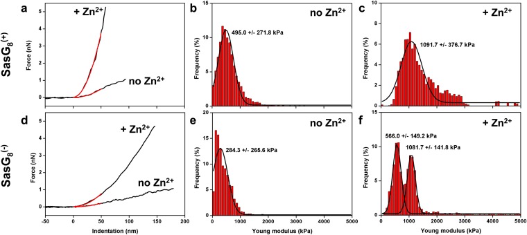
Staphylococcus aureus surface protein SasG promotes cell-cell adhesion during the accumulation phase of biofilm formation, but the molecular basis of this interaction remains poorly understood. Here, we unravel the mechanical properties of SasG on the surface of living bacteria, that is, in its native cellular environment. Nanoscale multiparametric imaging of living bacteria reveals that Zn(2+) strongly increases cell wall rigidity and activates the adhesive function of SasG. Single-cell force measurements show that SasG mediates cell-cell adhesion via specific Zn(2+)-dependent homophilic bonds between β-sheet-rich G5-E domains on neighboring cells. The force required to unfold individual domains is remarkably strong, up to ∼500 pN, thus explaining how SasG can withstand physiological shear forces. We also observe that SasG forms homophilic bonds with the structurally related accumulation-associated protein of Staphylococcus epidermidis, suggesting the possibility of multispecies biofilms during host colonization and infection. Collectively, our findings support a model in which zinc plays a dual role in activating cell-cell adhesion: adsorption of zinc ions to the bacterial cell surface increases cell wall cohesion and favors the projection of elongated SasG proteins away from the cell surface, thereby enabling zinc-dependent homophilic bonds between opposing cells. This work demonstrates an unexpected relationship between mechanics and adhesion in a staphylococcal surface protein, which may represent a general mechanism among bacterial pathogens for activating cell association.

Keywords: SasG; Staphylococcus aureus; adhesion; atomic force microscopy; biofilms.


Conflict of interest statement

The authors declare no conflict of interest.

Figures

Fig. 1.
Fig. 1.
Role of SasG in cell–cell adhesion. (A) Schematic representation of the SasG structure emphasizing the A domain, not engaged in cell–cell adhesion, and the B repeat sequence containing G5 domains (78 residues) in a tandem array, separated by the E regions (50 residues). (BE) Optical microscopy images of S. aureus cells expressing full-length SasG [SasG8(+) cells] after resuspension in TBS buffer (B) or in TBS buffer containing 1 mM of ZnCl2 (C), after addition of 1 mM EDTA (D), and further addition of 1 mM ZnCl2 (E). (F and G) Control experiment using S. aureus expressing no SasG [SasG(−) cells] in TBS buffer (F) or in TBS containing 1 mM of ZnCl2 (G).
Fig. 2.
Fig. 2.
Nanoscale multiparametric imaging unravels the structural and biophysical properties of S. aureus. (A) Height image of two dividing S. aureus cells expressing SasG [SasG8(+)] in TBS buffer and simultaneous (E) Young modulus and (I) adhesion image. Data were obtained in the same conditions following addition of 1 mM ZnCl2 (B, F, and J), of 1 mM ZnCl2 then 1 mM of EDTA (C, G, and K), and following further addition of 1 mM of ZnCl2 (D, H, and L). Control experiment using S. aureus cells expressing no SasG [SasG8(−)] in TBS buffer (M, O, and Q) or in TBS containing 1 mM ZnCl2 (N, P, and R). (Scale bars: 1 µm.) Similar results were obtained for five SasG8(+) cells and five SasG8(−) cells from different cultures.
Fig. S1.
Fig. S1.
Zn2+ induces major changes in cell surface structure. (A, D, G, and J) High-resolution height images and (B, E, H, and K) corresponding deflection images obtained for SasG8(+) cells (A, B, D, and E) and SasG8(−) cells (G, H, J, and K) in TBS in the absence (A, B, G, and H) or presence (D, E, J, and K) of Zn2+. (C, F, I, and L) Cross-sections taken along the white lines of the height images. (Scale bars: 100 nm.) Similar results were obtained on five different SasG8(+) cells and SasG8(−) cells.
Fig. S2.
Fig. S2.
Cell wall mechanics is strongly influenced by Zn2+. (A and D) Representative force-indentation curves (black lines) obtained on 500-nm × 500-nm areas of S. aureus cells expressing SasG [A, SasG8(+)] or S. aureus cells expressing no SasG [D, SasG8(−)] in TBS buffer containing Zn2+ or not; shown in red are the fits obtained with the Hertz model on the first 50 nm. (B, C, E, and F) Histograms of Young modulus values obtained for SasG8(+) cells (B and C) and SasG8(−) cells (E and F) in TBS in the absence (B and E) or presence (C and F) of Zn2+. For each condition, data from a total of n = 3,072 curves from three different cells are shown.
Fig. S3.
Fig. S3.
Cell surface properties are not influenced by Ca2+. (A and B) Height images of four S. aureus cells expressing SasG [SasG8(+)] in TBS buffer (A) or in TBS buffer with 1 mM of CaCl2 (B), and simultaneous (C and D) Young modulus and (E and F) adhesion images. (Scale bars: 1 µm.) Similar results were obtained using multiple cells from different cultures. (G and H) Adhesion force histograms obtained in TBS buffer (G) or in TBS buffer with 1 mM of CaCl2 (H) between two SasG8(+) cells. Similar results were obtained with multiple cells from different cultures.
Fig. S4.
Fig. S4.
Single-molecule imaging reveals that Zn2+ induces SasG exposure on the cell surface. (A and B) Adhesion force maps (500 nm × 500 nm) (Scale bar: 100 nm.) obtained by recording force curves across the surface of SasG8(+) cells in TBS buffer (A) or in TBS buffer with 1 mM of ZnCl2 (B) using antibody-labeled tips. (Insets) Maps from independent experiments. (C) Representative force curves from the maps in B, documenting the detection and unfolding of single SasG proteins only when zinc is present. (D–F) Results obtained by performing the same analyses on SasG8(−) cells. Similar data were obtained in three independent experiments using different tips and cell cultures.
Fig. 3.
Fig. 3.
Single-cell force spectroscopy of SasG bonds. (A) Adhesion force and (B) rupture distance histograms obtained in TBS buffer for three pairs (see three different colors) of S. aureus cells expressing full-length SasG [SasG8(+)]. “n.a.” values are the percentages of nonadhesive events. Inset in B is a fluorescence image of a single bacterium attached to the colloidal probe. (C–H) Force data obtained in the same conditions following addition of 1 mM of ZnCl2 (C and D), of 1 mM of ZnCl2, then 1 mM of EDTA (E and F), and following further addition of 1 mM of ZnCl2 (G and H). Insets in D and H show representative force signatures and their occurrence (n = 1,200 curves in each condition). All curves were obtained using a contact time of 0.1 s, an applied force of 250 pN, and an approach and retraction speed of 1.0 µm/s.
Fig. 4.
Fig. 4.
Mechanical strength of SasG domains. (A) Representative force curve displaying sawtooth profiles with up to eight low force peaks followed by eight high force peaks, that were well fitted with the extensible worm-like-chain (WLC) model using a persistence length lp of 0.4 nm (blue and red lines). (B) Unfolding force and (C) contour length gain (ΔLc) histograms documenting the sequential unfolding of E domains (blue; n = 51), followed by the more stable G5 domains (red; n = 58).
Fig. S5.
Fig. S5.
Intercellular adhesion involves Zn2+-dependent homophilic bonds between SasG multidomains. (A) Adhesion force and (B) rupture distance histograms with representative curves obtained for a pair of S. aureus cells expressing no SasG [SasG8(−)] in TBS buffer with 1 mM of ZnCl2. The “n.a.” stands for nonadhesive events. (C and D) Force data obtained for a SasG8(+)–SasG8(−) pair in TBS buffer with 1 mM of ZnCl2. (E and F) Force data obtained for a pair of S. aureus cells expressing only one G5–E segment [SasG1(+)] in TBS buffer with 1 mM of ZnCl2. All curves were obtained using a contact time of 0.1 s, an applied force of 250 pN, and an approach and retraction speed of 1.0 µm/s. (G and H) Simultaneous height (G) and adhesion (H) images of a SasG1(+) cell in TBS buffer with 1 mM of ZnCl2 recorded using multiparametric imaging with a silicon nitride tip. (Scale bars: 1 µm.) For each condition, similar results were obtained for five cell pairs (A–F) or five cells (G and H) from different cultures.
Fig. S6.
Fig. S6.
SasG forms homophilic bonds with the structurally related Aap protein. (A and C) Adhesion force and (B and D) rupture distance histograms with representative curves obtained between a S. aureus cell expressing SasG [SasG8(+)] and a S. epidermidis cell expressing Aap [Aap(+)], in TBS buffer in the absence (A and B) or presence (C and D) of 1 mM of ZnCl2. (EH) Force data for the interaction between a S. aureus cell expressing no SasG [SasG8(−)] and a S. epidermidis cell expressing Aap [Aap(+)]. For each condition, similar results were obtained for five cell pairs from three different cultures.
Fig. 5.
Fig. 5.
Proposed model for the zinc-dependent activation of intercellular adhesion. (A) Zn2+ alters the cell wall organization thereby making SasG fully functional. In the absence of Zn2+, cell wall components including negatively charged teichoic acids (TAs) and poly-γ-glutamic acid (PGA), are freely distributed and protrude from the cell surface, therefore masking SasG proteins. Addition of Zn2+ dramatically alters the cell wall organization by interaction with negatively charged TA and PGA. Zn2+ binding increases the cell surface stiffness and decreases the cell surface roughness, allowing SasG proteins to be fully available for interaction. (B) Zn2+ promotes homophilic bonds between protruding SasG repeats on opposing cells, thereby leading to intercellular adhesion. For the sake of clarity the A domain is not shown.

References

    1. Costerton JW, Stewart PS, Greenberg EP. Bacterial biofilms: A common cause of persistent infections. Science. 1999;284(5418):1318–1322. - PubMed
    1. López D, Vlamakis H, Kolter R. Biofilms. Cold Spring Harb Perspect Biol. 2010;2(7):a000398. - PMC - PubMed
    1. Lister JL, Horswill AR. Staphylococcus aureus biofilms: Recent developments in biofilm dispersal. Front Cell Infect Microbiol. 2014;4:178. - PMC - PubMed
    1. Hogan S, Stevens NT, Humphreys H, O’Gara JP, O’Neill E. Current and future approaches to the prevention and treatment of staphylococcal medical device-related infections. Curr Pharm Des. 2015;21(1):100–113. - PubMed
    1. Hobley L, Harkins C, MacPhee CE, Stanley-Wall NR. Giving structure to the biofilm matrix: An overview of individual strategies and emerging common themes. FEMS Microbiol Rev. 2015;39(5):649–669. - PMC - PubMed

Publication types

MeSH terms

LinkOut - more resources