. 2015 Dec;22(6):e493-7.
doi: 10.3747/co.22.2542.

Isolated brain metastases as first site of recurrence in prostate cancer: case report and review of the literature

Affiliations

Isolated brain metastases as first site of recurrence in prostate cancer: case report and review of the literature

J Craig et al. Curr Oncol. 2015 Dec.

Abstract

Fewer than 2% of patients with metastatic prostate cancer (pca) develop brain metastases. Autopsy series have confirmed the rarity of brain metastases. When present, brain metastases occur in end stage, once the pca is castrate-resistant and spread to other sites is extensive. Here, we present a rare case of a patient with pca who developed a solitary parenchymal brain metastasis as first site of relapse 9 years after radical therapy. The patient underwent craniotomy and excision of the tumour. A second recurrence was also isolated to the brain. In the literature, pca patients with brain metastases have a poor mean survival of 1-7.6 months. The patient in our case report experienced a relatively favourable outcome, surviving 19 months after his initial brain relapse.

Keywords: Prostate cancer; brain metastasis; recurrence.


Figures

FIGURE 1
FIGURE 1
Intra-axial enhancing mass lesion measuring 3.5 cm (anterior–posterior) by 3.1 cm (transverse) by 2.9 cm (craniocaudal) in the right frontal lobe, with surrounding vasogenic edema and areas of necrosis.
FIGURE 2
FIGURE 2
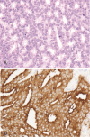
The tumour (A) consisted of a proliferation of relatively monomorphous epithelial cells disposed in small glandular configurations, consistent with moderately differentiated metastatic adenocarcinoma (hematoxylin and eosin stain, 400× original magnification), and (B) showed intense diffuse immunoreactivity for prostate-specific antigen (PSA) (immunohistochemistry for PSA, 400× original magnification).
FIGURE 3
FIGURE 3
Axial T1 post-gadolinium magnetic resonance image, 1 month after right frontal craniotomy. The surgical cavity demonstrates blood products and minimal peripheral enhancement. Resolution of the surrounding vasogenic edema is evident. No residual tumour and no other enhancing brain lesions are observed.
FIGURE 4
FIGURE 4
Significant disease progression, with multiple dura-based enhancing lesions, vasogenic edema, and a mild midline shift toward the left, is seen. The brain parenchyma is involved by the largest exophytic lesion in the right middle cranial fossa.

References

    1. Howlader N, Noone AM, Krapcho M, et al., editors. SEER Cancer Statistics Review, 1975–2010. Bethesda, MD: US National Cancer Institute; 2013.
    1. Tremont-Lukats IW, Bobustuc G, Lagos GK, Lolas K, Kyritsis AP, Puduvalli VK. Brain metastasis from prostate carcinoma: the M.D. Anderson Cancer Center experience. Cancer. 2003;98:363–8. doi: 10.1002/cncr.11522. - DOI - PubMed
    1. Baumann MA, Holoye PY, Choi H. Adenocarcinoma of prostate presenting as brain metastasis. Cancer. 1984;54:1723–5. doi: 10.1002/1097-0142(19841015)54:8<1723::AID-CNCR2820540840>3.0.CO;2-G. - DOI - PubMed
    1. Saitoh H, Hida M, Shimbo T, Nakamura K, Yamagata J, Satoh T. Metastatic patterns of prostatic cancer. Correlation between sites and number of organs involved. Cancer. 1984;54:3078–84. doi: 10.1002/1097-0142(19841215)54:12<3078::AID-CNCR2820541245>3.0.CO;2-U. - DOI - PubMed
    1. Benjamin R. Neurologic complications of prostate cancer. Am Fam Physician. 2002;65:1834–40. - PubMed

LinkOut - more resources