

Rotator cuff tears: An evidence based approach
- PMID: 26716086
- PMCID: PMC4686437
- DOI: 10.5312/wjo.v6.i11.902
Rotator cuff tears: An evidence based approach
Abstract
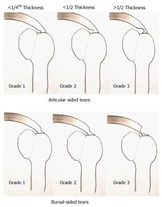
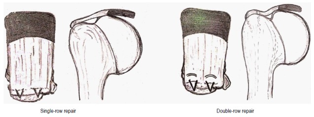
Lesions of the rotator cuff (RC) are a common occurrence affecting millions of people across all parts of the globe. RC tears are also rampantly prevalent with an age-dependent increase in numbers. Other associated factors include a history of trauma, limb dominance, contralateral shoulder, smoking-status, hypercholesterolemia, posture and occupational dispositions. The challenge lies in early diagnosis since a high proportion of patients are asymptomatic. Pain and decreasing shoulder power and function should alert the heedful practitioner in recognizing promptly the onset or aggravation of existing RC tears. Partial-thickness tears (PTT) can be bursal-sided or articular-sided tears. Over the course of time, PTT enlarge and propagate into full-thickness tears (FTT) and develop distinct chronic pathological changes due to muscle retraction, fatty infiltration and muscle atrophy. These lead to a reduction in tendon elasticity and viability. Eventually, the glenohumeral joint experiences a series of degenerative alterations - cuff tear arthropathy. To avert this, a vigilant clinician must utilize and corroborate clinical skill and radiological findings to identify tear progression. Modern radio-diagnostic means of ultrasonography and magnetic resonance imaging provide excellent visualization of structural details and are crucial in determining further course of action for these patients. Physical therapy along with activity modifications, anti-inflammatory and analgesic medications form the pillars of nonoperative treatment. Elderly patients with minimal functional demands can be managed conservatively and reassessed at frequent intervals. Regular monitoring helps in isolating patients who require surgical interventions. Early surgery should be considered in younger, active and symptomatic, healthy patients. In addition to being cost-effective, this helps in providing a functional shoulder with a stable cuff. An easily reproducible technique of maximal strength and sturdiness should by chosen among the armamentarium of the shoulder surgeon. Grade 1 PTTs do well with debridement while more severe lesions mandate repair either by trans-tendon technique or repair following conversion into FTT. Early repair of repairable FTT can avoid appearance and progression of disability and weakness. The choice of surgery varies from surgeon-to-surgeon with arthroscopy taking the lead in the current scenario. The double-row repairs have an edge over the single-row technique in some patients especially those with massive tears. Stronger, cost-effective and improved functional scores can be obtained by the former. Both early and delayed postoperative rehabilitation programmes have led to comparable outcomes. Guarded results may be anticipated in patients in extremes of age, presence of comorbidities and severe tear patters. Overall, satisfactory results are obtained with timely diagnosis and execution of the appropriate treatment modality.
Keywords: Double row repair; Full thickness tear; Healing; Magnetic resonance imaging; Natural history; Partial thickness tears; Rotator cuff tears; Single row repair; Ultrasonography.
Figures
References
-
- Park SE, Panchal K, Jeong JJ, Kim YY, Kim JH, Lee JY, Ji JH. Intratendinous rotator cuff tears: prevalence and clinical and radiological outcomes of arthroscopically confirmed intratendinous tears at midterm follow-up. Am J Sports Med. 2015;43:415–422. - PubMed
-
- Yamaguchi K, Ditsios K, Middleton WD, Hildebolt CF, Galatz LM, Teefey SA. The demographic and morphological features of rotator cuff disease. A comparison of asymptomatic and symptomatic shoulders. J Bone Joint Surg Am. 2006;88:1699–1704. - PubMed
Publication types
LinkOut - more resources
Full Text Sources
Other Literature Sources
