

Iatrogenic perforation of esophagus successfully treated with Endoscopic Vacuum Therapy (EVT)
- PMID: 26716109
- PMCID: PMC4683128
- DOI: 10.1055/s-0034-1392566
Iatrogenic perforation of esophagus successfully treated with Endoscopic Vacuum Therapy (EVT)
Abstract
Background and study aims: Endoscopic Vacuum Therapy (EVT) has been reported as a novel treatment option for esophageal leakage. We present our results in the treatment of iatrogenic perforation with EVT in a case series of 10 patients.
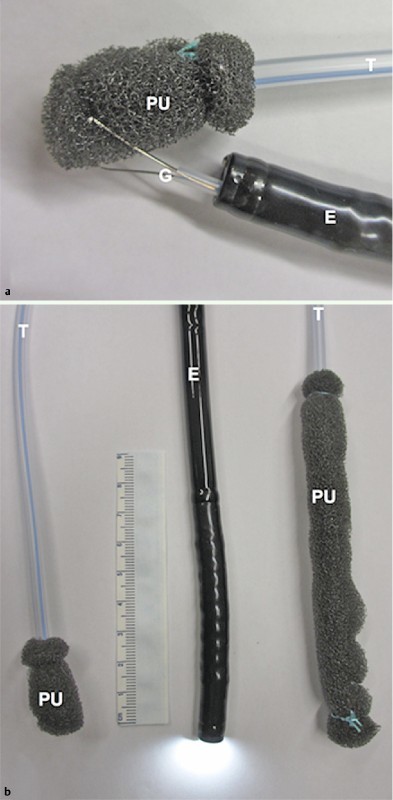
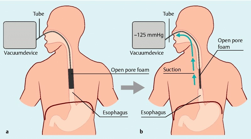
Patients and methods: An open pore polyurethane drainage was placed either intracavitary through the perforation defect or intraluminal covering the defect zone. Application of vacuum suction with an electronic device (continuous negative pressure, -125 mmHg) resulted in defect closure and internal drainage.
Results: Esophageal perforations were located from the cricopharyngeus (4/10) to the esophagogastric junction (2/10). EVT was feasible in all patients. Eight patients were treated with intraluminal EVT, one with intracavitary EVT, and one with both types of treatments. All perforations (100 %) were healed in within a median of (3 - 7) days. No stenosis occurred, no complications were observed, and no additional operative treatment was necessary.
Conclusions: Our study suggests that intraluminal EVT will play an important role in endoscopic management of esophageal perforation.
Conflict of interest statement
Figures

References
-
- Carrott P W Jr, Low D E. Advances in the management of esophageal perforation. Thorac Surg Clin. 2011;21:541–555. - PubMed
-
- Schmidt S C, Strauch S, Rosch T. et al.Management of esophageal perforations. Surg Endosc. 2010;24:2809–2813. - PubMed
-
- Schorsch T, Muller C, Loske G. Endoscopic vacuum therapy of anastomotic leakage and iatrogenic perforation in the esophagus. Surg Endosc. 2013;27:2040–2045. - PubMed
LinkOut - more resources
Full Text Sources
Other Literature Sources