

Tea nanoparticle, a safe and biocompatible nanocarrier, greatly potentiates the anticancer activity of doxorubicin
- PMID: 26716507
- PMCID: PMC4868728
- DOI: 10.18632/oncotarget.6711
Tea nanoparticle, a safe and biocompatible nanocarrier, greatly potentiates the anticancer activity of doxorubicin
Abstract
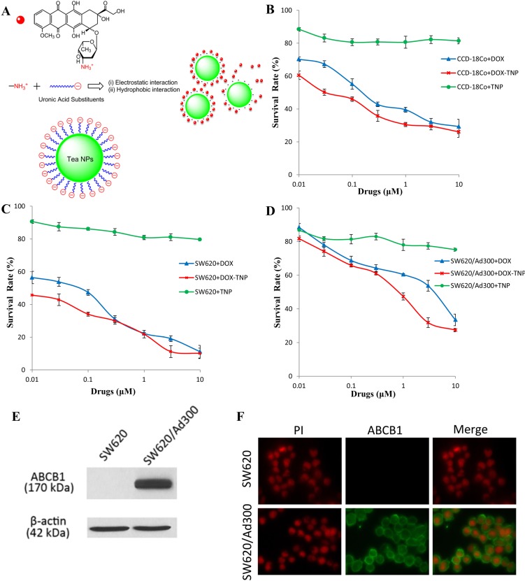
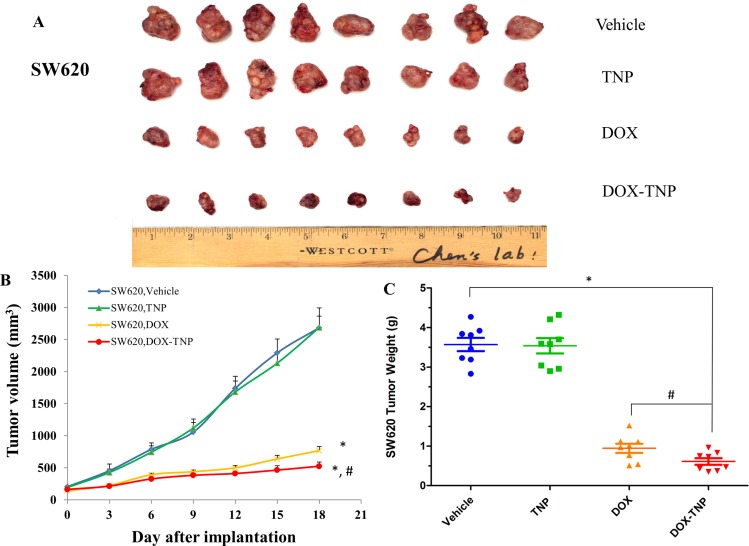
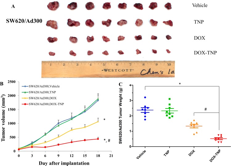
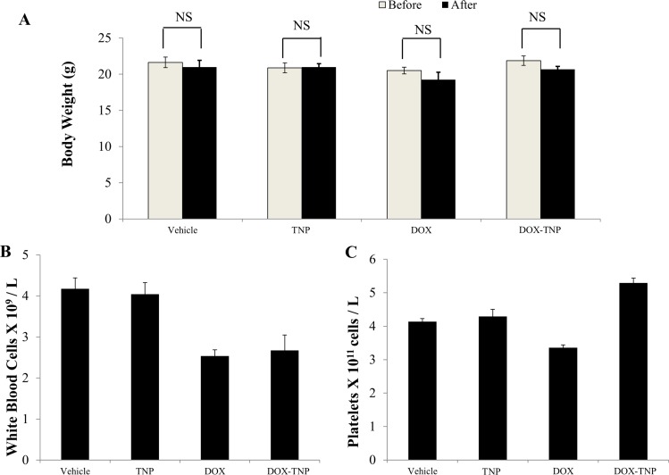
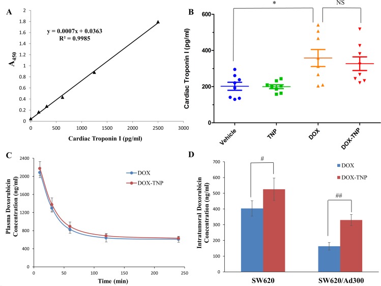
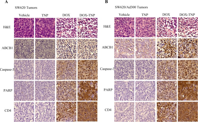
An infusion-dialysis based procedure has been developed as an approach to isolate organic nanoparticles from green tea. Tea nanoparticle (TNP) can effectively load doxorubicin (DOX) via electrostatic and hydrophobic interactions. We established an ABCB1 overexpressing tumor xenograft mouse model to investigate whether TNP can effectively deliver DOX into tumors and bypass the efflux function of the ABCB1 transporter, thereby increasing the intratumoral accumulation of DOX and potentiating the anticancer activity of DOX. MTT assays suggested that DOX-TNP showed higher cytotoxicity toward CCD-18Co, SW620 and SW620/Ad300 cells than DOX. Animal study revealed that DOX-TNP resulted in greater inhibitory effects on the growth of SW620 and SW620/Ad300 tumors than DOX. In pharmacokinetics study, DOX-TNP greatly increased the SW620 and SW620/Ad300 intratumoral concentrations of DOX. But DOX-TNP had no effect on the plasma concentrations of DOX. Furthermore, TNP is a safe nanocarrier with excellent biocompatibility and minimal toxicity. Ex vivo IHC analysis of SW620 and SW620/Ad300 tumor sections revealed evidence of prominent antitumor activity of DOX-TNP. In conclusion, our findings suggested that natural nanomaterials could be useful in combating multidrug resistance (MDR) in cancer cells and potentiating the anticancer activity of chemotherapeutic agents in cancer treatment.
Keywords: biocompatibility; drug delivery; multidrug resistance; tea nanoparticle; tumor xenograft.
Conflict of interest statement
The authors have declared no potential conflicts of interest.
Figures
Similar articles
-
Surfactin-based nanoparticles loaded with doxorubicin to overcome multidrug resistance in cancers.Int J Nanomedicine. 2018 Mar 21;13:1723-1736. doi: 10.2147/IJN.S157368. eCollection 2018. Int J Nanomedicine. 2018. PMID: 29606866 Free PMC article.
-
Immuno-oncology agent IPI-549 is a modulator of P-glycoprotein (P-gp, MDR1, ABCB1)-mediated multidrug resistance (MDR) in cancer: In vitro and in vivo.Cancer Lett. 2019 Feb 1;442:91-103. doi: 10.1016/j.canlet.2018.10.020. Epub 2018 Nov 1. Cancer Lett. 2019. PMID: 30391357 Free PMC article.
-
Functional Doxorubicin-Loaded Omega-3 Unsaturated Fatty Acids Nanoparticles in Reversing Hepatocellular Carcinoma Multidrug Resistance.Med Sci Monit. 2021 Feb 1;27:e927727. doi: 10.12659/MSM.927727. Med Sci Monit. 2021. PMID: 33524008 Free PMC article.
-
Structure-Based Design, Synthesis, and Biological Evaluation of New Triazolo[1,5-a]Pyrimidine Derivatives as Highly Potent and Orally Active ABCB1 Modulators.J Med Chem. 2020 Dec 24;63(24):15979-15996. doi: 10.1021/acs.jmedchem.0c01741. Epub 2020 Dec 5. J Med Chem. 2020. PMID: 33280384
-
Reversal effect of FW-04-806, a macrolide dilactone compound, on multidrug resistance mediated by ABCB1 and ABCG2 in vitro and in vivo.Cell Commun Signal. 2019 Sep 1;17(1):110. doi: 10.1186/s12964-019-0408-5. Cell Commun Signal. 2019. PMID: 31472682 Free PMC article.
Cited by
-
Lipid-Saporin Nanoparticles for the Intracellular Delivery of Cytotoxic Protein to Overcome ABC Transporter-Mediated Multidrug Resistance In Vitro and In Vivo.Cancers (Basel). 2020 Feb 21;12(2):498. doi: 10.3390/cancers12020498. Cancers (Basel). 2020. PMID: 32098067 Free PMC article.
-
Natural Nano-Drug Delivery System in Coptidis Rhizoma Extract with Modified Berberine Hydrochloride Pharmacokinetics.Int J Nanomedicine. 2021 Sep 14;16:6297-6311. doi: 10.2147/IJN.S323685. eCollection 2021. Int J Nanomedicine. 2021. PMID: 34552326 Free PMC article.
-
Bafetinib (INNO-406) reverses multidrug resistance by inhibiting the efflux function of ABCB1 and ABCG2 transporters.Sci Rep. 2016 May 9;6:25694. doi: 10.1038/srep25694. Sci Rep. 2016. PMID: 27157787 Free PMC article.
-
In Search of Panacea-Review of Recent Studies Concerning Nature-Derived Anticancer Agents.Nutrients. 2019 Jun 25;11(6):1426. doi: 10.3390/nu11061426. Nutrients. 2019. PMID: 31242602 Free PMC article. Review.
-
Dynamic Formation of Green Tea Cream and the Identification of Key Components Using the "Knock-Out/Knock-In" Method.Foods. 2023 Aug 8;12(16):2987. doi: 10.3390/foods12162987. Foods. 2023. PMID: 37627986 Free PMC article.
References
-
- Jemal A, Murray T, Ward E, Samuels A, Tiwari RC, Ghafoor A, Feuer EJ, Thun MJ. Cancer statistics, 2005. CA Cancer J Clin. 2005;55:10–30. - PubMed
-
- Cockerill GS, Lackey KE. Small molecule inhibitors of the class 1 receptor tyrosine kinase family. Curr Top Med Chem. 2002;2:1001–1010. - PubMed
-
- Gottesman MM. Mechanisms of cancer drug resistance. Annu Rev Med. 2002;53:615–627. - PubMed
-
- Deeley RG, Westlake C, Cole SP. Transmembrane transport of endo- and xenobiotics by mammalian ATP-binding cassette multidrug resistance proteins. Physiol Rev. 2006;86:849–899. - PubMed
Publication types
MeSH terms
Substances
Grants and funding
LinkOut - more resources
Full Text Sources
Other Literature Sources
