. 2015 Dec 30;10(12):e0145550.
doi: 10.1371/journal.pone.0145550. eCollection 2015.

The Development of Macrophage-Mediated Cell Therapy to Improve Skeletal Muscle Function after Injury

Affiliations

The Development of Macrophage-Mediated Cell Therapy to Improve Skeletal Muscle Function after Injury

Viktoriya Rybalko et al. PLoS One. .

Abstract

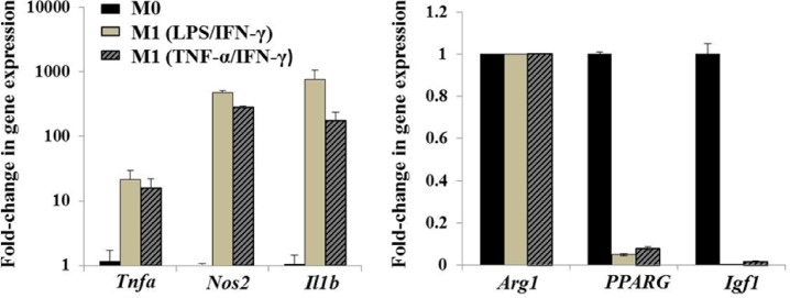
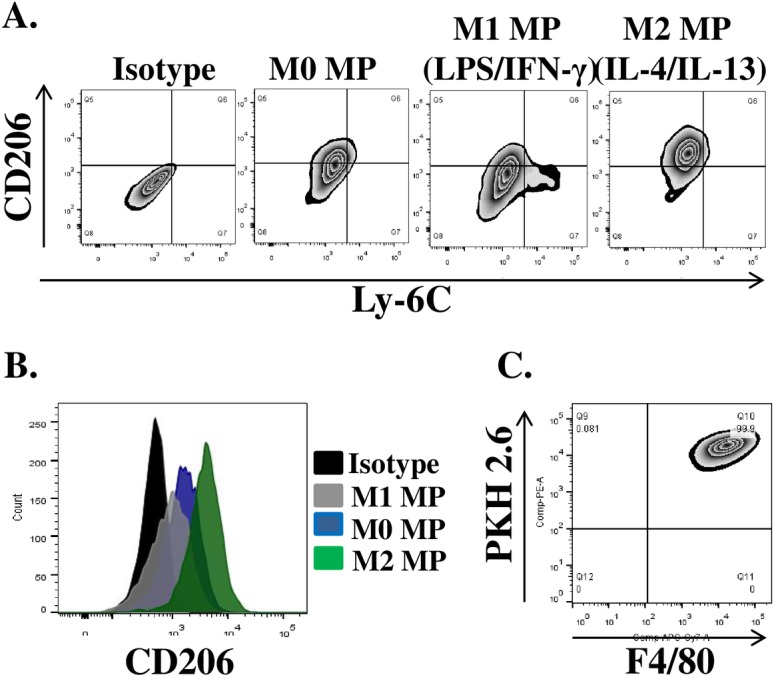
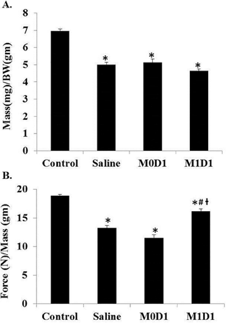
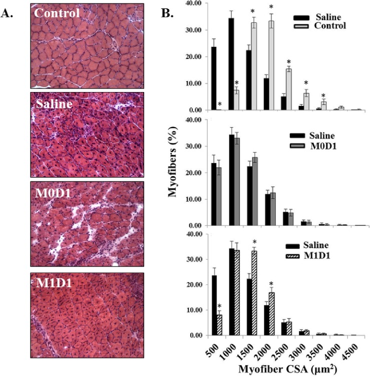
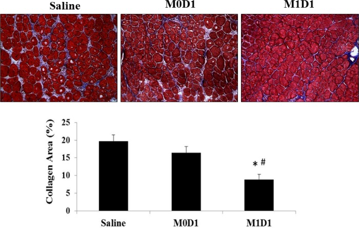
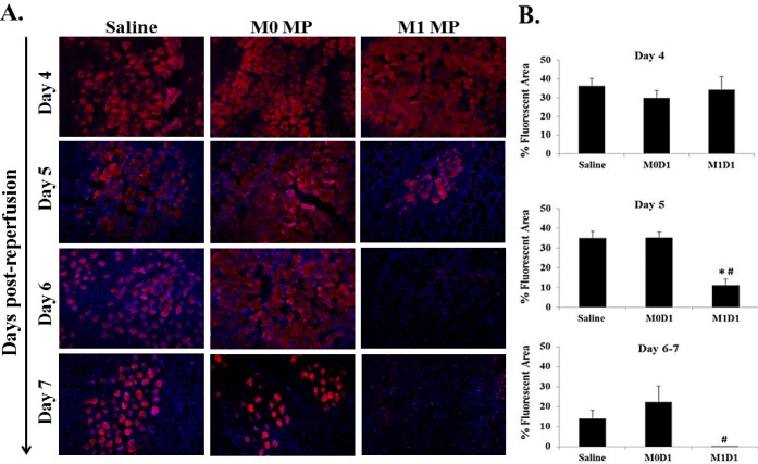
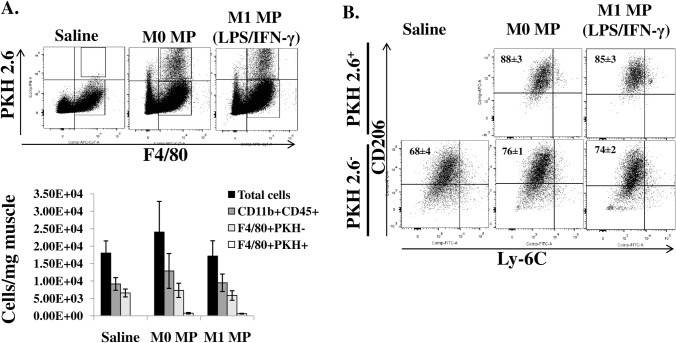
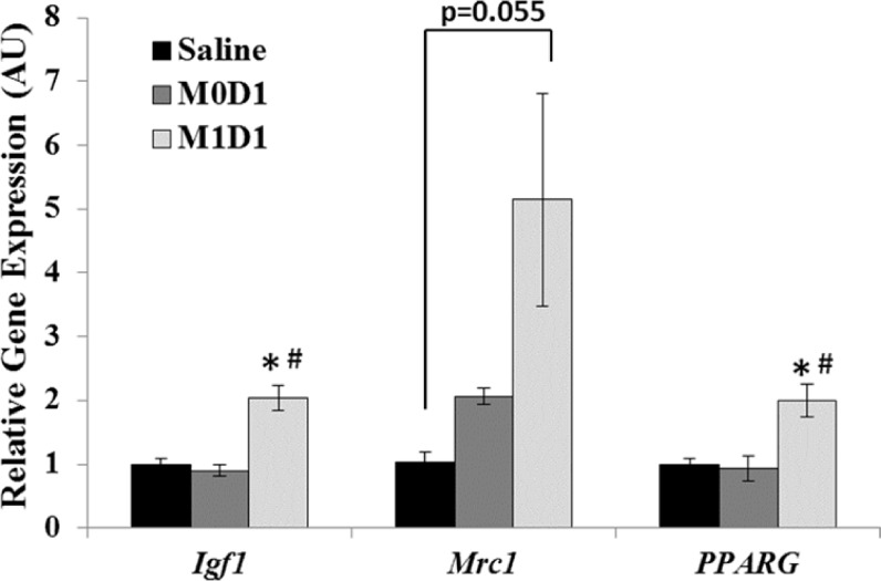
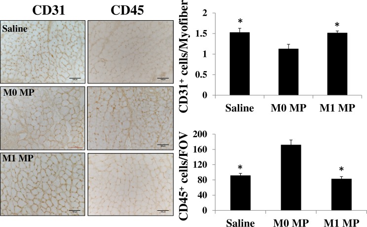
Skeletal muscle regeneration following acute injury is a multi-step process involving complex changes in tissue microenvironment. Macrophages (MPs) are one of the key cell types involved in orchestration and modulation of the repair process. Multiple studies highlight the essential role of MPs in the control of the myogenic program and inflammatory response during skeletal muscle regeneration. A variety of MP phenotypes have been identified and characterized in vitro as well as in vivo. As such, MPs hold great promise for cell-based therapies in the field of regenerative medicine. In this study we used bone-marrow derived in vitro LPS/IFN-y-induced M1 MPs to enhance functional muscle recovery after tourniquet-induced ischemia/reperfusion injury (TK-I/R). We detected a 15% improvement in specific tension and force normalized to mass after M1 (LPS/IFN-γ) MP transplantation 24 hours post-reperfusion. Interestingly, we found that M0 bone marrow-derived unpolarized MPs significantly impaired muscle function highlighting the complexity of temporally coordinated skeletal muscle regenerative program. Furthermore, we show that delivery of M1 (LPS/IFN-γ) MPs early in regeneration accelerates myofiber repair, decreases fibrotic tissue deposition and increases whole muscle IGF-I expression.


Conflict of interest statement

Competing Interests: The authors have declared that no competing interests exist.

Figures

Fig 1
Fig 1. In vitro polarization of bone marrow-derived macrophages.
BM MPs were either left untreated (M0) or treated with LPS/IFN-γ or TNF-α/IFN-γ for 42 hours to induce classical (M1) activation phenotype. Real-time PCR was performed to evaluate inflammatory (Tnfa, Nos2, Il1b) and anti-inflammatory (Arg1, PPARG, Igf1) gene expression of MPs polarized with LPS/IFN-γ or TNF-α/IFN-γ (10 ng/ml) relative to M0 MPs. β-actin was used as internal calibrator gene. Values expressed as mean ± SD.
Fig 2
Fig 2. Phenotypic analysis of in vitro polarized BM MPs.
MPs were either left untreated (M0) or stimulated with 10ng/ml LPS/IFN-γ and IL-4/IL-13for 42 hours to induce classical (M1) and alternative (M2) activation phenotypes respectively. Flow cytometry was used to evaluate the expression of CD206 and Ly-6C surface proteins. (A) Representative plots of surface protein after in vitro MP polarization, (B) Mean fluorescence intensity of CD206 expression on the surface of polarized MPs, (C) Representative plot of PKH2.6 label and F4/80expression by in vitro polarized macrophages prior to transplantation.
Fig 3
Fig 3. Gastrocnemius muscle mass normalized to body weight and force recovery 14 days post-reperfusion after saline injection or the delivery of 2x10⁶ in vitro polarized macrophages 24 h after TK-I/R injury.
(A) GAS mass(mg)/body weight (BW)(gm); (B) Force (N)/GAS mass (mg). Control n = 17, Saline n = 7, M0D1 (un-polarized) MPs n = 5, M1D1 (LPS/IFN-γ polarized (42h)) MPs n = 6. Values expressed as mean ± SEM. (*) p<0.05 relative to contralateral control; (#) p<0.05 relative to saline; (ƚ) p<0.05 relative to M0D1; one-way ANOVA, Tukey-HSD post-hoc.
Fig 4
Fig 4. Analysis of the myofiber distribution 14 days post-reperfusion in GAS muscles treated with saline or 2x10⁶ in vitro polarized macrophages 24h after TK-I/R injury.
(A) Representative H&E images of uninjured control GAS and injured, saline and MP treated GAS, at 14 days post-reperfusion; (B) Top: myofiber distribution in saline-treated group (black bars) relative to the contralateral uninjured control GAS(white bars); Middle: myofiber distribution after saline treatment (black bars) compared to M0 MP injected GAS (grey bars); Bottom: myofiber distribution following saline treatment (black bars) compared to M1(LPS/IFN-γ) MP treated GAS (pattern fill bars). n = 5/group; 3 fields of view/animal. Values expressed as mean ± SEM; (*) p<0.05 relative to saline; Student’s t-test.
Fig 5
Fig 5. Trichrome staining for the evaluation of collagen deposition in gastrocnemius muscles 14 days post-reperfusion treated with saline or 2x10⁶ in vitro polarized macrophages 24h after TK-I/R injury.
(*) p<0.05 compared to saline; (#) p<0.05 compared to M0D1; n = 3/group; 3 fields of view/animal; values expressed as mean ± SEM; one-way ANOVA, Tukey-HSD post-hoc.
Fig 6
Fig 6. Evan’s Blue dye staining of damaged myofibers at 4, 5, 6 and 7 days post reperfusion in muscles treated with saline or 2x10⁶ M0 and M1 macrophages 24 h after TK-I/R injury.
(A) Representative images of EBD+ staining (Day 4 (n = 3); Day 5 (n = 3); Day 6 (n = 1); Day 7 (n = 1)). Damaged fibers (EBD permeable) represented in red, DAPI (blue) used to counterstain cell nuclei; (B) Measurements of percent (%) fluorescent area. Days 4 and 5 post-I/R, n = 3/group, except D4-M0D1 n = 2; 3 fields of view/animal. Days 6–7, n = 2, 3 fields of view/animal. Values expressed as mean ± SEM. (*) p<0.05 compared to saline; (#) p<0.05 compared to M0D1; one-way ANOVA, Tukey-HSD post-hoc.
Fig 7
Fig 7. In vivo quantification and characterization of MP populations 5 days after TK-I/R injury.
A) Flow cytometric identification and quantification of myeloid and MP cell populations in TK-injured muscle. Total myeloid cells (CD45+CD11b+), resident MPs (CD11b+F4/80+PKH2.6-), transplanted MPs (CD11b+F4/80+PKH2.6+).Values expressed as mean ±SEM, n = 3, B) Expression of CD206 and Ly-6C surface proteins on F4/80+MP populations in control and MP-treated muscles. Values expressed as mean ±SEM, n = 3.
Fig 8
Fig 8. Real-Time PCR evaluation of gene expression in gastrocnemius muscles 5 days post-reperfusion treated with saline or 2x10⁶ M0 or M1 macrophages 24h after TK-I/R injury.
Gene expression of IGF-I (Igf1), CD206 (Mrc1) and PPAR-γ (PPARG) relative to saline. Values expressed as mean transcript values ± SEM; n = 3. 3- phosphoglycerate kinase (Pgk1) was used as internal reference gene. (*) p<0.05 compared to saline; (#) p<0.05 compared to M0D1, one-way ANOVA, Tukey-HSD post-hoc.
Fig 9
Fig 9. Assessment of tissue revascularization and immune cell infiltrate in saline or MP-treated TK-I/R injured muscles 14 days post-reperfusion.
Representative images of D14 regenerating skeletal muscle stained with anti-CD31antibody identifying vascular endothelium and anti-CD45 staining for immune cells, scale bar 100μm. Quantification graphs of the number of CD31+ cell per myofiber (capillary/fiber ratio) and CD45+ cells per field of view (FOV). M0 group n = 3, M1 and saline groups n = 4–5, 3–5 images/muscle, values presented as mean ± SEM, (*) p<0.05 relative to M0 MP group

References

    1. Ten Broek RW, Grefte S, Von den Hoff JW. Regulatory factors and cell populations involved in skeletal muscle regeneration. Journal of cellular physiology. 2010;224(1):7–16. 10.1002/jcp.22127 . - DOI - PubMed
    1. Tidball JG. Mechanisms of muscle injury, repair, and regeneration. Comprehensive Physiology. 2011;1(4):2029–62. 10.1002/cphy.c100092 . - DOI - PubMed
    1. Nathan C. Points of control in inflammation. Nature. 2002;420(6917):846–52. 10.1038/nature01320 . - DOI - PubMed
    1. Blaisdell FW. The pathophysiology of skeletal muscle ischemia and the reperfusion syndrome: a review. Cardiovascular surgery. 2002;10(6):620–30. . - PubMed
    1. Chen XK, Rathbone CR, Walters TJ. Treatment of tourniquet-induced ischemia reperfusion injury with muscle progenitor cells. The Journal of surgical research. 2011;170(1):e65–73. 10.1016/j.jss.2011.05.061 . - DOI - PubMed

Publication types

MeSH terms