. 2016 Feb;13(2):1517-24.
doi: 10.3892/mmr.2015.4726. Epub 2015 Dec 28.

Bone marrow-derived mesenchymal stem cells rescue injured H9c2 cells via transferring intact mitochondria through tunneling nanotubes in an in vitro simulated ischemia/reperfusion model

Affiliations

Bone marrow-derived mesenchymal stem cells rescue injured H9c2 cells via transferring intact mitochondria through tunneling nanotubes in an in vitro simulated ischemia/reperfusion model

Hui Han et al. Mol Med Rep. 2016 Feb.

Abstract

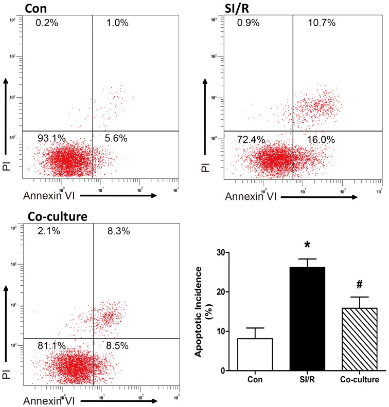
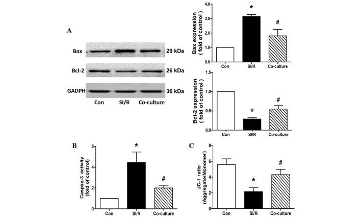
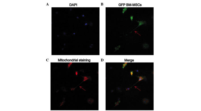
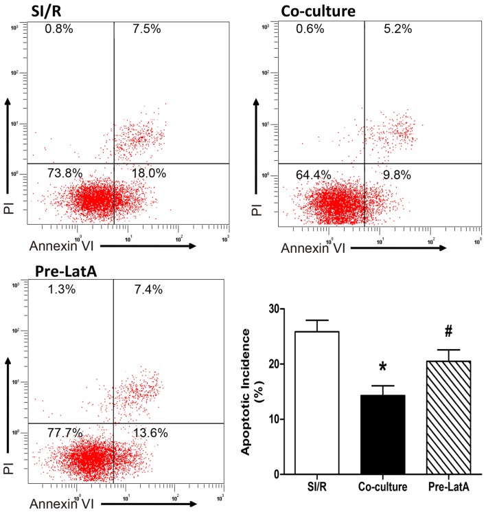
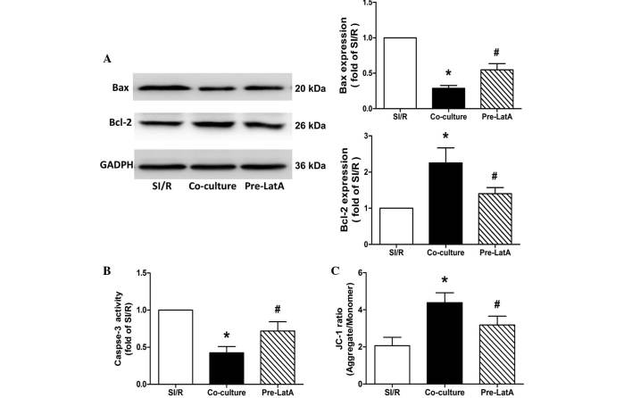
The transplantation of mesenchymal stem cells (MSCs) is considered to be a promising treatment for ischemic heart disease; however, the therapeutic effects and underlying mechanisms of action require further evaluation. Mitochondrial dysfunction is a key event in simulated ischemia/reperfusion (SI/R) injury. The purpose of the present study was to investigate the mechanism of mitochondrial transfer, which may be involved the antiapoptotic action of co-culture with MSCs. An in vitro model of simulated ischemia/reperfusion (SI/R) was used in the present study. The apoptotic indexes were significantly increased when H9c2 cardiomyocytes were induced in the SI/R group. Following co-culture with bone marrow-derived (BM)-MSCs, H9c2 cells exhibited marked resistance against the SI/R-induced apoptotic process. Besides, mitochondrial transfer via a tunneling nanotube (TNT) like structure was detected by confocal fluorescent microscopy. In addition, following pretreated with latrunculin-A (LatA), an inhibitor of TNT formation, the BM-MSCs were not able to rescue injured H9c2 cells from apoptosis, as previously observed. In conclusion, the anti-apoptotic ability of BM-MSCs may be partially attributed to the recovery of mitochondrial dysfunction in SI/R, and the formation of TNTs appears to be involved in this action of mitochondrial transfer between adjacent cells.


Figures

Figure 1
Figure 1
Identification of isolated BM-MSCs. Flow cytometric analysis of (A) CD90 (B) CD45, (C) CD29 and (D) CD31. The results showed >90% of the BM-MSCs were positive for CD90 or CD29, whereas <3% of the BM-MSCs were positive for CD45 or CD31. BM-MSCs, bone marrow-derived mesenchymal stem cells; CD, cluster of differentiation.
Figure 2
Figure 2
Effects of co-culture with BM-MSCs on apoptosis in the control (Con), SI/R and SI/R co-cultured with BM-MSCs (Co-culture) groups. (A) Changes in apoptotic incidence in the Con, SI/R and Co-culture groups. Data are presented as the mean ± standard deviation. *P<0.05, vs. Con; #P<0.05, vs. SI/R. BM-MSCs, bone marrow-derived mesenchymal stem cells; SI/R, simulated ischemia/reperfusion.
Figure 3
Figure 3
Effects of co-culture with BM-MSCs on apoptosis and mitochondrial membrane potential in the control (Con), SI/R and SI/R co-cultured with BM-MSCs (Co-culture) groups. (A) Representative western blots showing the changes in the expression Bax and Bcl-2 in H9c2 cells from the Con, SI/R and Co-culture groups. (B) Changes in caspase-3 activity in H9c2 cells from the Con, SI/R and Co-culture groups. (C) Changes in mitochondrial membrane potential in H9c2 cells from the Con, SI/R and Co-culture groups. Data are presented as the mean ± standard deviation (*P<0.05, vs. Con; #P<0.05, vs. SI/R). BM-MSCs, bone marrow-derived mesenchymal stem cells; SI/R, simulated ischemia/reperfusion; Bcl-2, B cell lymphoma-2; Bax, Bcl-2-associated X protein; JC-1, 5,5′,6,6′-tetrachloro-1,1′,3,3′-tetraethylbenzimidazole-carbocyanide iodine.
Figure 4
Figure 4
Mitochondria transfer following direct co-culture of ischemia/reperfusion-stimulated H9c2 cells with BM-MSCs. Arrows indicate the TNT-like structures. (A) Blue nucleus stained by DAPI; (B) Green fluorescence from GFP BM-MSCs; (C) Red mitochondria stained by MitoTracker Deep Red; (D) Merge of A–C. The arrows indicate the TNT-like structures. SI/R, simulated ischemia/reperfusion; BM-MSCs, bone marrow-derived mesenchymal stem cells; GFP, green fluorescence protein. GFP, green fluorescence protein; BM-MSCs, bone marrow-derived mesenchymal stem cells.
Figure 5
Figure 5
Evaluation of apoptotic incidence in the SI/R, Co-culture and Pre-LatA groups. Data are presented as the mean ± standard deviation (*P<0.05, vs. SI/R. #P<0.05, vs. Co-culture). BM-MSCs, bone marrow-derived mesenchymal stem cells; SI/R, simulated ischemia/reperfusion; Co-culture, SI/R co-cultured with BM-MSCs; LatA, latrunculin-A; Pre-LatA, pretreated with LatA.
Figure 6
Figure 6
Effects of co-culture with BM-MSCs on apoptosis and mitochondrial membrane potential in the SI/R, Co culture and Pre LatA groups. (A) Representative western blotting of the changes in the expression levels of Bax and Bcl-2 in H9c2 cells. (B) Changes in caspase-3 activity in H9c2 cells from the different groups. (C) Changes in mitochondrial membrane potential in H9c2 cells from the different groups. Data are presented as the mean ± standard deviation (*P<0.05, vs. SI/R; #P<0.05, vs. Co-culture). BM-MSCs, bone marrow-derived mesenchymal stem cells; SI/R, simulated ischemia/reperfusion; Co-culture, SI/R co-cultured with BM-MSCs; LatA, latrunculin-A; Pre-LatA, pretreated with LatA; Bcl-2, B cell lymphoma-2; Bax, Bcl-2-associated X protein; JC-1, 5,5′,6,6′-tetrachloro-1,1′,3,3′-tetraethylbenzimidazole-carbocyanide iodine.

References

    1. Roger VL, Go AS, Lloyd-Jones DM, Adams RJ, Berry JD, Brown TM, Carnethon MR, Dai S, de Simone G, Ford ES, et al. Heart disease and stroke statistics-2011 update: A report from the American heart association. Circulation. 2011;123:e18–e209. doi: 10.1161/CIR.0b013e3182009701. - DOI - PMC - PubMed
    1. Forouzanfar MH, Moran AE, Flaxman AD, Roth G, Mensah GA, Ezzati M, Naghavi M, Murray CJ. Assessing the global burden of ischemic heart disease, part 2: analytic methods and estimates of the global epidemiology of ischemic heart disease in 2010. Glob Heart. 2012;7:331–342. doi: 10.1016/j.gheart.2012.10.003. - DOI - PMC - PubMed
    1. Yu D, Li M, Tian Y, Liu J, Shang J. Luteolin inhibits ROS-activated MAPK pathway in myocardial ischemia/reperfusion injury. Life Sci. 2015;122:15–25. doi: 10.1016/j.lfs.2014.11.014. - DOI - PubMed
    1. Chen C, Feng Y, Zou L, Wang L, Chen HH, Cai JY, Xu JM, Sosnovik DE, Chao W. Role of extracellular RNA and TLR3-Trif signaling in myocardial ischemia-reperfusion injury. J Am Heart Assoc. 2014;3:e000683. doi: 10.1161/JAHA.113.000683. - DOI - PMC - PubMed
    1. Tanaka-Esposito C, Chen Q, Lesnefsky EJ. Blockade of electron transport before ischemia protects mitochondria and decreases myocardial injury during reperfusion in aged rat hearts. Transl Res. 2012;160:207–216. doi: 10.1016/j.trsl.2012.01.024. - DOI - PMC - PubMed

Publication types

MeSH terms