. 2016 Mar;64(3):168-78.
doi: 10.1369/0022155415626765. Epub 2015 Dec 30.

An Improved Immunostaining and Imaging Methodology to Determine Cell and Protein Distributions within the Bone Environment

Affiliations

An Improved Immunostaining and Imaging Methodology to Determine Cell and Protein Distributions within the Bone Environment

Hemanth Akkiraju et al. J Histochem Cytochem. 2016 Mar.

Abstract

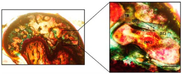
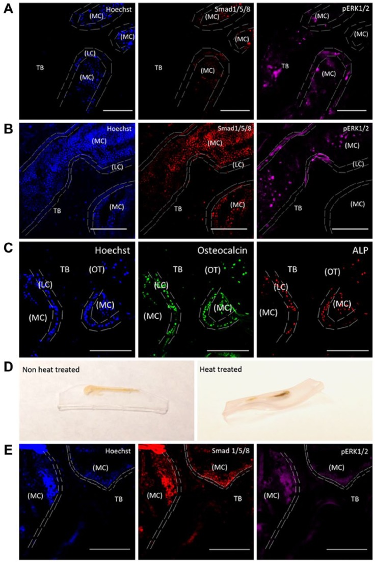
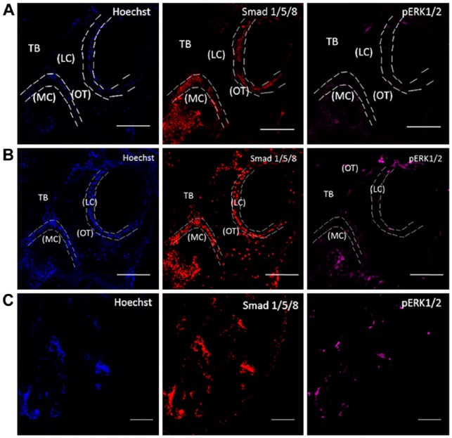
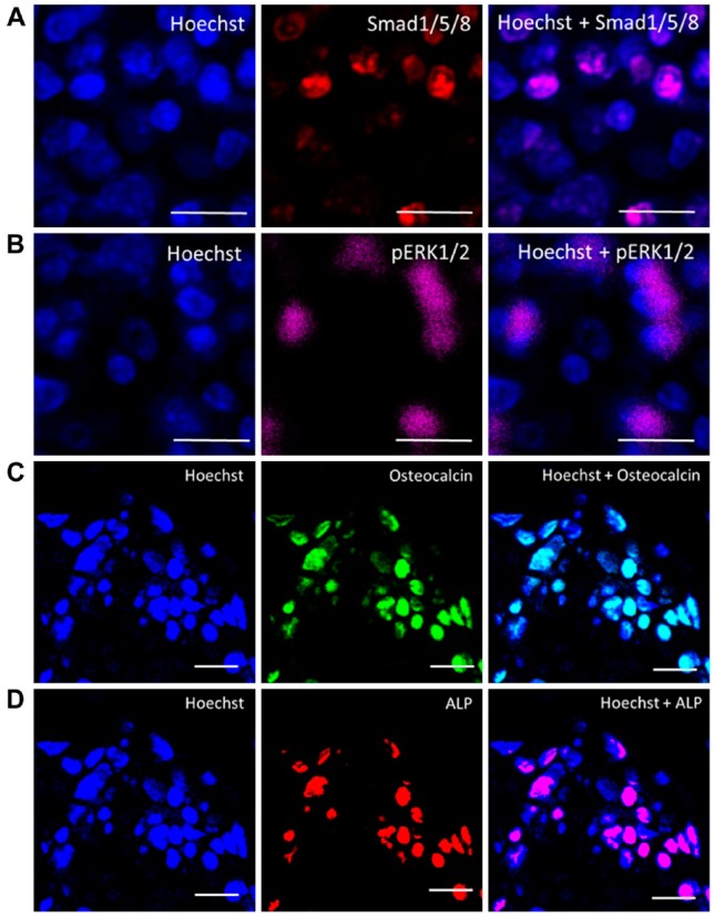
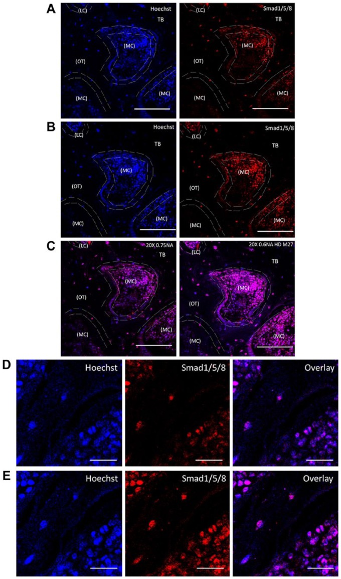
Bone is a dynamic tissue that undergoes multiple changes throughout its lifetime. Its maintenance requires a tight regulation between the cells embedded within the bone matrix, and an imbalance among these cells may lead to bone diseases such as osteoporosis. Identifying cell populations and their proteins within bone is necessary for understanding bone biology. Immunolabeling is one approach used to visualize proteins in tissues. Efficient immunolabeling of bone samples often requires decalcification, which may lead to changes in the structural morphology of the bone. Recently, methyl-methacrylate embedding of non-decalcified tissue followed by heat-induced antigen retrieval has been used to process bone sections for immunolabeling. However, this technique is applicable for bone slices below 50-µm thickness while fixed on slides. Additionally, enhancing epitope exposure for immunolabeling is still a challenge. Moreover, imaging bone cells within the bone environment using standard confocal microscopy is difficult. Here we demonstrate for the first time an improved methodology for immunolabeling non-decalcified bone using a testicular hyaluronidase enzyme-based antigen retrieval technique followed by two-photon fluorescence laser microscopy (TPLM) imaging. This procedure allowed us to image key intracellular proteins in bone cells while preserving the structural morphology of the cells and the bone.

Keywords: bone; confocal microscopy; immunolabeling; osteoblasts; osteocytes; testicular hyaluronidase; two photon fluorescence laser microscopy.


Conflict of interest statement

Competing Interests: The authors declared no potential competing interest with respect to the research, authorship, and/or publication of this article.

Figures

Figure 1.
Figure 1.
Trabecular morphology of the MMA-embedded femur sample. Femurs from mice were processed in MMA and stained by Villanueva Osteochrome. Phase-contrast images of the samples show osteocytes (OT) represented as dark brown dense osteoid seams of trabecular bone (TB; green) and osteoblasts or lining cells (LC) of the marrow cavity (MC; light red). Sections were imaged in the cancellous or TB of the mouse femur around the MC where the LC or the active osteoblasts reside alongside of OTs (indicated by arrows). Images were taken using 20× magnification. Scale, 50 µm. Imaging was conducted using a Nikon TMS (model TMS-F #211153).
Figure 2.
Figure 2.
Testicular hyaluronidase-based antigen retrieval of MMA-embedded femur slices. MMA-embedded samples were immunostained for Smad 1/5/8, pERK1/2, osteocalcin and alkaline phosphatase (ALP). Hoechst staining was used to identify cells within the bone (blue). Heat-induced antigen retrieval was used as a comparison. Sections were imaged in the cancellous or the trabecular bone (TB) of the mouse femur around the marrow cavity (MC) where the lining cells (LC) or the active osteoblasts reside alongside osteocytes (OT). (A) MMA-embedded samples not treated with testicular hyaluronidase and imaged using TPLM. Tissue sections were stained with Hoechst (blue), regions of bone growth and proteins associated with bone cell activity were stained with Smad 1/5/8 (red) and pERK1/2 (magenta). (B) MMA samples treated with testicular hyaluronidase and stained as in (A) were imaged using TPLM. (C) MMA samples treated with testicular hyaluronidase and stained for osteocalcin (green), and ALP (red). (D) Heat-induced antigen retrieval caused morphing of the thick MMA-embedded bone sample if not mounted onto a permanent slide fixture. (E) MMA samples were heat treated and imaged using TPLM. Images were stained with Hoechst (blue), Smad 1/5/8 (red) and pERK1/2 (magenta). All images were taken using 20× magnification. Scale, 100 µM.
Figure 3.
Figure 3.
Two-photon excitation laser microscopy imaging of MMA-embedded samples. Imaging of MMA-embedded samples stained with Hoechst (blue), Smad 1/5/8 (red) and pERK1/2 (magenta). (A) Conventional confocal setup. The collected images were of low-resolution and demonstrated high autofluorescence. (B) Two-photon excitation laser microscopy imaging. Images show nuclei and labeling for proteins. (C) A representative tile scan image that was used to examine the entire sample section. Sections were imaged in the cancellous or trabecular bone (TB) of the mouse femur around the marrow cavity (MC) where the lining cells (LC) or active osteoblasts reside alongside osteocytes (OT). Images were taken at 20× magnification. Scale (A, B) 100 µm; (C) 200 µm.
Figure 4.
Figure 4.
High-resolution imaging of cells expressing Smad1/5/8, p-ERK, osteocalcin and alkaline phosphatase. Magnified regions of the images were taken using a 20× objective with 2× magnification at 4096 × 4096 pixels. (A) Smad1/5/8 (red), Hoechst (blue). (B) pERK1/2 (magenta). (C) Osteocalcin (green). (D) ALP (red) and Hoechst. The overlay images at high resolution clearly distinguish the different expressions of these proteins within the cells. Scale, 10 µm.
Figure 5.
Figure 5.
Objective differences and their impact on sample brightness. MMA-embedded bone samples were TPLM-imaged on an inverted microscope Zeiss 780 using (A) 20×/NA 0.75 designed to be imaged with coverslips or a (B) 20×/NA 0.6 HD M27 for samples without coverslips. The minor differences in spatial resolution are compared based on the working distances of each objective. For the samples without coverslips, an objective of 20×/NA 0.6 HD M27 had a working distance of 1.7 mm, which provided the best imaging conditions for a better field of view, as shown in the overlay image in (C). Sections were imaged in the cancellous or trabecular bone (TB) of the mouse femur around the marrow cavity (MC) where the lining cells (LC) or the active osteoblasts reside alongside osteocytes (OT), labeled for Smad1/5/8 (red) and Hoechst (blue). bar representing 100 µm. High magnified images clearly distinguish objective differences in the staining of Smad (red) and Hoechst (blue) with overlay in images taken using (D) 20×/0.75NA, and (E) 20×/0.6NA HD M27. Images taken at 20× magnification. Scale (C) 100 µm; (D) 20 µm.

References

    1. Akkiraju H, Bonor J, Olli K, Bowen C, Bragdon B, Coombs H, Donahue LR, Duncan R, Nohe A. (2015). Systemic injection of CK2.3, a novel peptide acting downstream of bone morphogenetic protein receptor BMPRIa, leads to increased trabecular bone mass. J Orthop Res 33:208-215. - PMC - PubMed
    1. An YH, Martin KL. (2003). Histological techniques for decalcified bone and cartilage Handbook of Histology Methods for Bone and Cartilage. New York: Humana Press.
    1. Bancroft JD, Stevens A. (1982). Theory and Practice of Histological Techniques, 2nd edition, New York: Churchill Livingstone.
    1. Blythe D, Hand NM, Jackson P, Barrans SL, Bradbury RD, Jack AS. (1997). Use of methyl methacrylate resin for embedding bone marrow trephine biopsy specimens. J Clin Pathol 50:45-49. - PMC - PubMed
    1. Brandi ML. (2009). Microarchitecture, the key to bone quality. Rheumatology (Oxford) 48 Suppl 4:iv3-8. - PubMed

Publication types

LinkOut - more resources