. 2016 Feb 1;196(3):1412-8.
doi: 10.4049/jimmunol.1501886. Epub 2015 Dec 30.

Complement Component 3 Is Regulated by TWIST1 and Mediates Epithelial-Mesenchymal Transition

Affiliations

Complement Component 3 Is Regulated by TWIST1 and Mediates Epithelial-Mesenchymal Transition

Min Soon Cho et al. J Immunol. .

Abstract

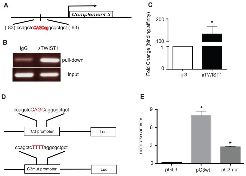
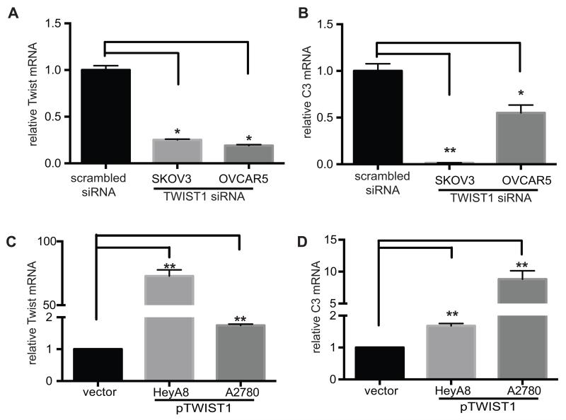
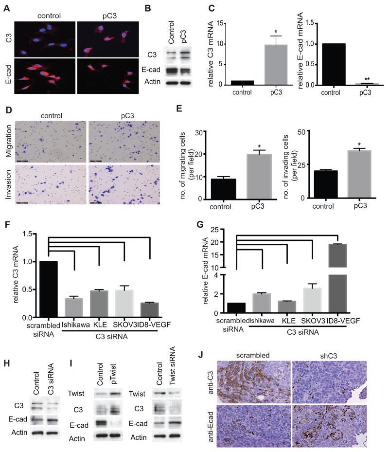
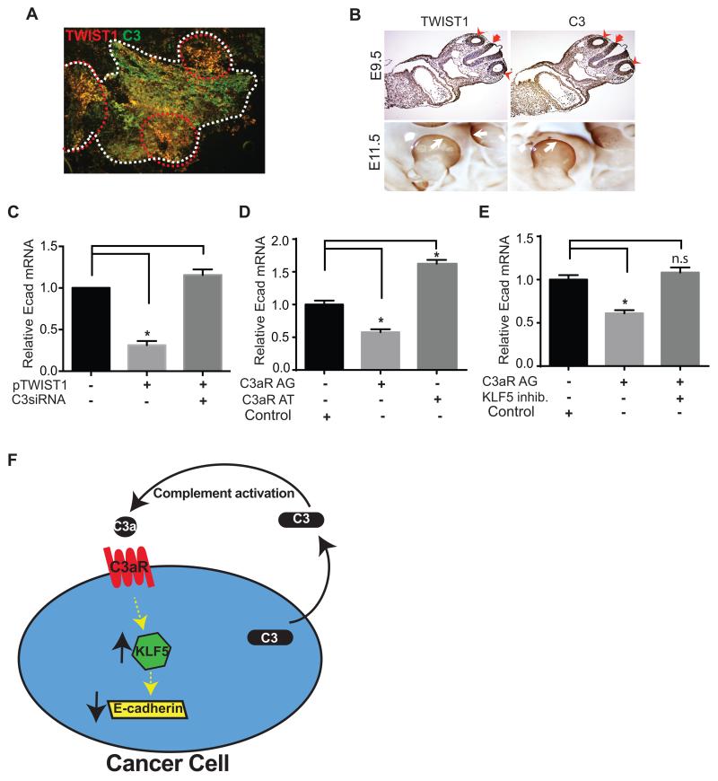
We have previously shown that complement component 3 (C3) is secreted by malignant epithelial cells. To understand the mechanism of upregulation of C3 expression in tumor cells, we studied the C3 promoter and identified that twist basic helix-loop-helix transcription factor 1 (TWIST1) binds to the C3 promoter and enhances its expression. Because TWIST1 mediates epithelial-mesenchymal transition (EMT), we studied the effect of C3 on EMT and found that C3 decreased E-cadherin expression on cancer cells and promoted EMT. We showed that C3-induced reduction in E-cadherin expression in ovarian cancer cells was mediated by C3a and is Krüppel-like factor 5 dependent. We investigated the association between TWIST1 and C3 in malignant tumors and in murine embryos. TWIST1 and C3 colocalized at the invasive tumor edges, and in the neural crest and limb buds of mouse embryos. Our results identified TWIST1 as a transcription factor that regulates C3 expression during pathologic and physiologic EMT.


Figures

FIGURE 1
FIGURE 1. TWIST1 binds to the C3 promoter
(A) A schematic representation of TWIST1 binding site on the C3 promoter (−63 to −83). (B) PCR-amplification products using primers encompassing TWIST1 binding site on DNA template prepared by ChIP with TWIST1 antibody or control non-immune IgG were detected by agarose gel electrophoresis and ethidium bromide staining. (C) Results of qRT-PCR using primers encompassing TWIST1 binding site on the DNA templates prepared by ChIP with TWIST1 antibody or control IgG (n=3, p= 0.0017). (D) A schematic representation of reporter constructs generated by cloning the luciferase gene 3′ to the C3 promoter with either wild type (C3wt) or mutant (C3mut) TWIST1 core binding site. Four nucleotides in the TWIST1 core binding site were altered in C3mut (CAGC → TTTT). (E) HeyA8 ovarian cancer cells were co-transfected with plasmid containing C3 promoter constructs (pC3wt or pC3mut), plasmid containing TWIST1 cDNA, and plasmid containing Renilla luciferase. Promoterless luciferase plasmid (pGL3) was used instead of plasmids with C3 promoter constructs in the control group. Luciferase activity decreased by 66% in HeyA8 cells co-transfected with pC3mut plasmid as compared to that with pC3wt (n=3, p=0.001)
FIGURE 2
FIGURE 2. TWIST1 expression positively correlates with C3 expression
(A) Downregulation of TWIST1 by transfecting human ovarian cancer cells (SKOV3ip1 and OVCAR 5) with TWIST1 siRNAs was detected by measuring TWIST1 mRNA. (B) Subsequent to TWIST1 knockdown in C3-high SKOV3ip1 and OVCAR 5 cells, C3 expression was measured by quantitative RT-PCR. TWIST1 knockdown resulted in 99% reduction in C3 expression (n=3, p=0.001). (C) TWIST1 overexpression in C3-low HeyA8 and A2780 ovarian cancer cells by stable transfection of plasmid encoding TWIST1 cDNA (pTWIST1) resulted in 2 folds and 75 folds increase in TWIST1 expression, respectively (n=3, p=0.0001). (D) C3 mRNA levels in HeyA8 and A2780 cells stably transfected with the TWIST1 cDNA was measured by quantitative RT-PCR. TWIST1 overexpression in HeyA8 and A2780 cells resulted in 1.7 and 8.8 folds increase in C3 expression, respectively (n=3, p<0.005)
FIGURE 3
FIGURE 3. C3 expression negatively correlates with E-cadherin expression in vitro and in vivo
(A) After stable transduction of lentivirus carrying C3 cDNA (pC3) into low-C3 HeyA8, C3 expression (A, upper panels) or E-cadherin expression (A, lower panels) was detected by immunofluorescence microscopy, (B) by Western-blotting or (C) by quantitative RT-PCR, respectively. HeyA8 cells stably transduced by pC3 showed increase in C3 expression (B, left panel, n=3, p=0.002) and reduction in E-cadherin expression (B, right panel, n=3, p=0.008). (D) The functional effect of C3 overexpression in HeyA8 cells was studied by in vitro migration and invasion assays, using HeyA8 cells stably transfected with pC3. (E) HeyA8 cells overexpressing C3 showed 2.3 folds increase in migration ability (E, left panel, n=15, p=0.001) and 1.8 folds increase in invasion ability (E, right panel, n=15, p=0.001) as compared with those transfected with an empty control plasmid. (F) Ovarian (SKOV3 and OVCAR5) and endometrial (Ishikawa and KLE) cancer cell lines were transfected with C3 siRNA. C3 mRNA level was decreased by 55-75% in ovarian cancer cells and by 52-60% in endometrial cancer cells (n=3, p=0.001) as compared to scrambled siRNA transfected cells. (G) After C3 knockdown E-cadherin mRNA increased by 2.5-19 folds in ovarian cancer cells and by 1.3-2 folds in endometrial cancer cells (n=3, p=0.001). Cell lines transfected with scrambled siRNA served as controls. (H) After transient transfection of C3 siRNA into C3-high SKOV3, C3 and E-cadherin protein expression was determined by Western- and immunoblotting. Scrambled siRNA was used as a control. (I) After stable transfection of HeyA8 cells with plasmid containing TWIST1 cDNA (pTWIST1) and transfection of SKOV3 cells with TWIST1 siRNA, protein expression of TWIST1, C3, and E-cadherin was determined by Western-blotting. (J) Expression of E-cadherin and C3 in sections of ovarian tumors resected from tumor-bearing mice were studied using immunofluorescence microscopy. Tumors induced by ID8-VEGF cells transduced by C3 shRNA showed reduced expression of C3 and increased expression of E-cadherin as compared to tumors induced by ID8-VEGF cells transduced by scrambled shRNA.
FIGURE 4
FIGURE 4. Coexpression of C3 and TWIST1 in implanted tumors in mice and in developing murine embryos
(A) Expression of TWIST1 and C3 in implanted ovarian tumor tissue was analyzed using immunofluorescence microscopy. ID8-VEGF cells-induced tumors were resected and immunostained with TWIST1 (red) and C3 (green) antibodies. Borders of a tumor nodule are shown using white broken lines. Co-expression of TWIST1 and C3 (yellow) was more prominent at the invasive edges of the tumor (circumscribed with red broken lines). (B) Mouse embryos were collected at 9.5dpc (upper panels) and 11.5dpc (whole mounted, lower panels), and immunohistochemistry was performed using anti-TWIST1 or anti-C3 antibody. TWIST1 and C3 were co-localized in the otocysts (red arrow head) and hindbrain (red arrow) at 9.5dpc embryo, and in the limb-bud (white arrow) at 11.5dpc embryos. (C) To study the effect of C3 on the TWIST1-mediated gene regulation, E-cadherin mRNA level was measured before and after transient transfection of C3 siRNA into TWIST1-overexpressing HeyA8 cells (stably transfected with plasmid encoding TWIST1 cDNA). Scrambled siRNAs were used as negative controls. TWIST1 overexpression was associated with 70% decrease in E-cadherin mRNA level (n=3, *p=0.0002). C3 knockdown restored E-cadherin expression in TWIST1 overexpressing cells. (D) Incubation of SKOV ip1 human ovarian cancer cells with a C3a-receptor agonist (C3aR AG) for 2 days decreased E-cadherin mRNA level by 43% ± 0.1, and incubation with a C3a-receptor antagonist (C3aR AT) increased E-cadherin expression by 62% ± 0.2 (n=3, p<0.0001). Scrambled peptide served as the control. (E) Inhibition of KLF5 abrogated the C3a-receptor agonist-induced E-cadherin suppression (n=3, p<0.0001 and n.s= not significant). Scrambled peptide served as the control. (F) The schematic representation of the role of C3 in a pro-tumor autocrine loop in ovarian cancer. Cleavage of C3 secreted by ovarian cancer cells into tumor microenvironment generates C3a that binds to C3a-receptor on cancer cells. Activation of C3a-receptor increases KLF5, and KLF5 reduces E-cadherin expression.

Similar articles

Cited by

References

    1. Mastellos D, Lambris JD. Complement: more than a ‘guard’ against invading pathogens? Trends Immunol. 2002;23:485–491. - PubMed
    1. McLin VA, Hu CH, Shah R, Jamrich M. Expression of complement components coincides with early patterning and organogenesis in Xenopus laevis. Int. J. Dev. Biol. 2008;52:1123–1133. - PubMed
    1. Carmona-Fontaine C, Theveneau E, Tzekou A, Tada M, Woods M, Page KM, Parsons M, Lambris JD, Mayor R. Complement fragment C3a controls mutual cell attraction during collective cell migration. Dev. Cell. 2011;21:1026–1037. - PMC - PubMed
    1. Strainic MG, Liu J, Huang D, An F, Lalli PN, Muqim N, Shapiro VS, Dubyak GR, Heeger PS, Medof ME. Locally produced complement fragments C5a and C3a provide both costimulatory and survival signals to naive CD4+ T cells. Immunity. 2008;28:425–435. - PMC - PubMed
    1. Raedler H, Yang M, Lalli PN, Medof ME, Heeger PS. Primed CD8(+) T-cell responses to allogeneic endothelial cells are controlled by local complement activation. Am. J. Transplant. 2009;9:1784–1795. - PubMed

Publication types

MeSH terms