. 2015 Dec 30:6:263.
doi: 10.1186/s13287-015-0254-3.

Allogeneic guinea pig mesenchymal stem cells ameliorate neurological changes in experimental colitis

Affiliations

Allogeneic guinea pig mesenchymal stem cells ameliorate neurological changes in experimental colitis

Rhian Stavely et al. Stem Cell Res Ther. .

Abstract

Background: The use of mesenchymal stem cells (MSCs) to treat inflammatory bowel disease (IBD) is of great interest because of their immunomodulatory properties. Damage to the enteric nervous system (ENS) is implicated in IBD pathophysiology and disease progression. The most commonly used model to study inflammation-induced changes to the ENS is 2,4,6-trinitrobenzene-sulfonate acid (TNBS)-induced colitis in guinea pigs; however, no studies using guinea pig MSCs in colitis have been performed. This study aims to isolate and characterise guinea pig MSCs and then test their therapeutic potential for the treatment of enteric neuropathy associated with intestinal inflammation.

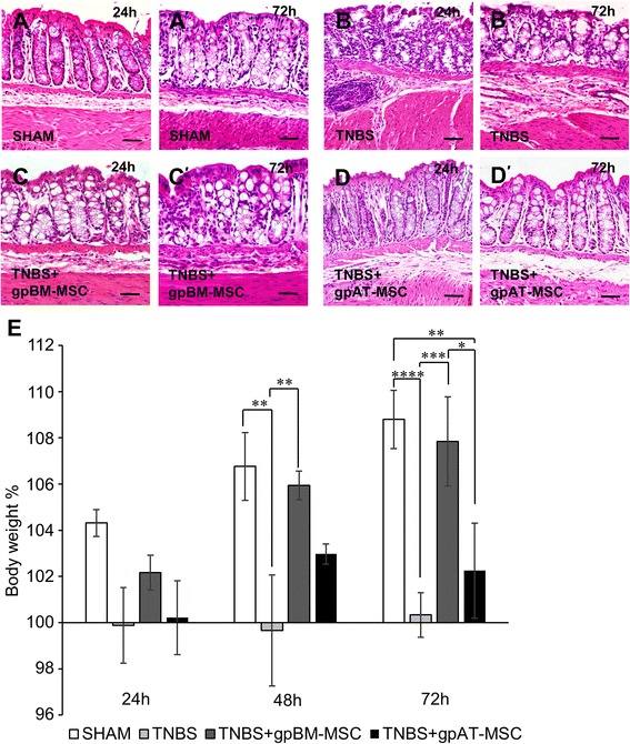
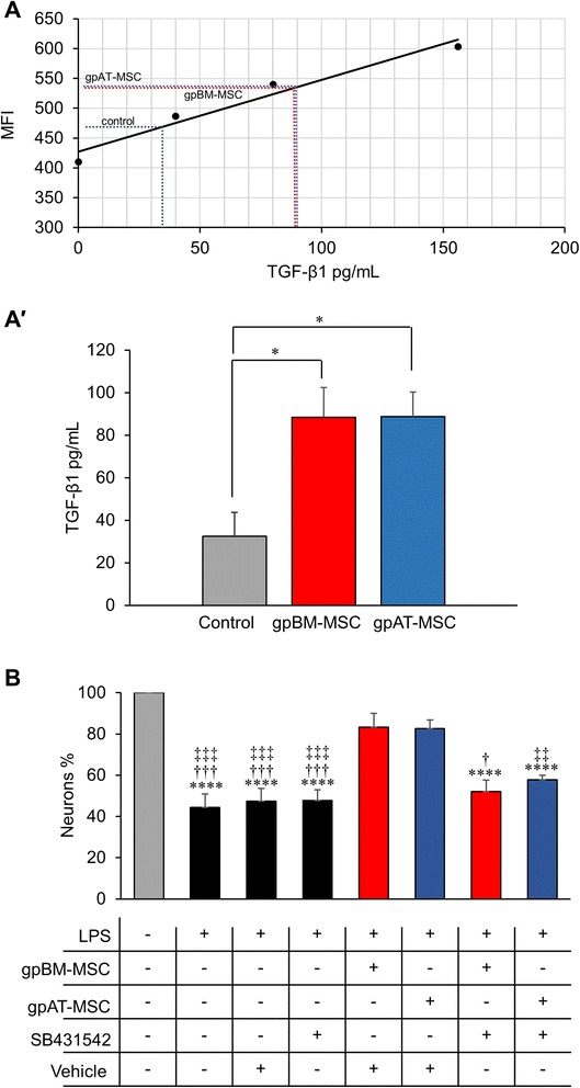
Methods: MSCs from guinea pig bone marrow and adipose tissue were isolated and characterised in vitro. In in vivo experiments, guinea pigs received either TNBS for the induction of colitis or sham treatment by enema. MSCs were administered at a dose of 1 × 10(6) cells via enema 3 h after the induction of colitis. Colon tissues were collected 24 and 72 h after TNBS administration to assess the level of inflammation and damage to the ENS. The secretion of transforming growth factor-β1 (TGF-β1) was analysed in MSC conditioned medium by flow cytometry.

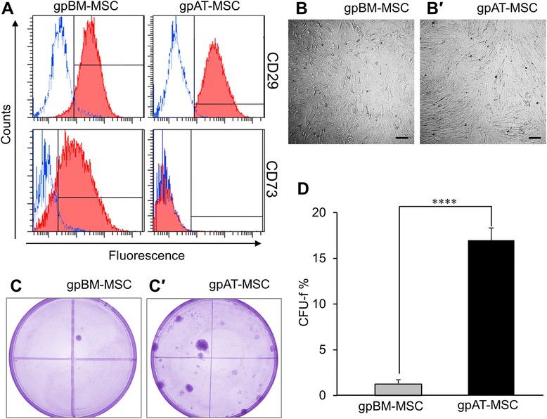
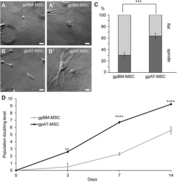
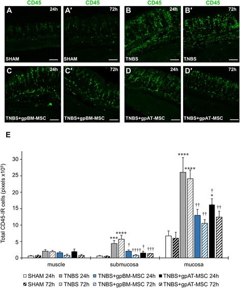
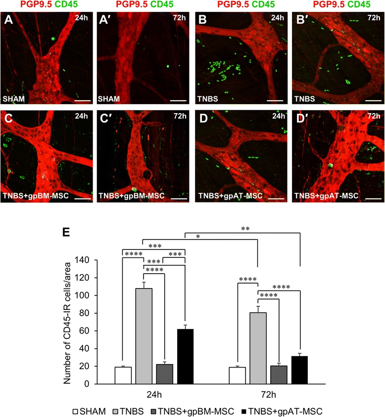
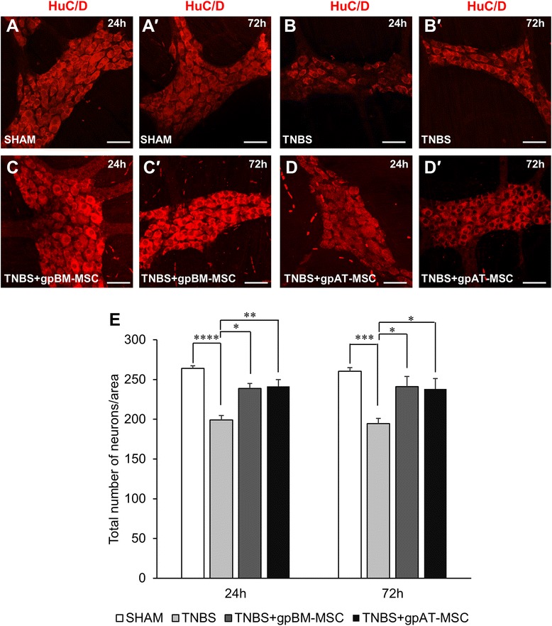
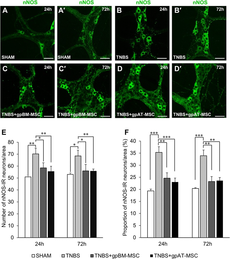
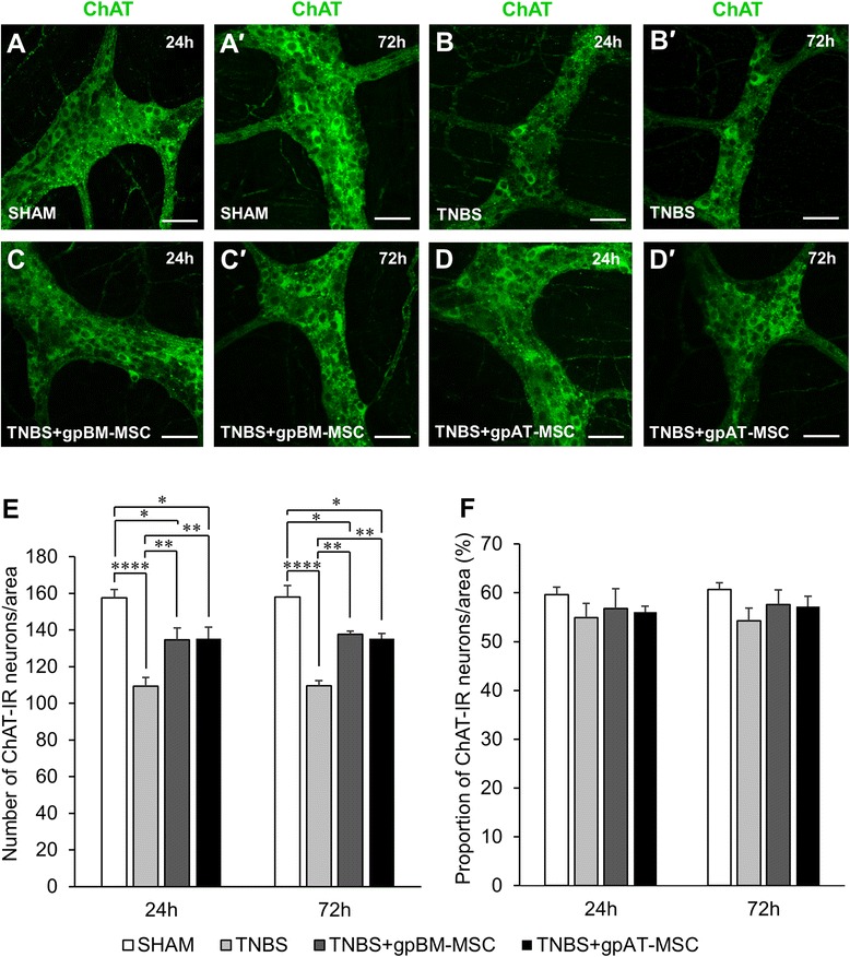
Results: Cells isolated from both sources were adherent to plastic, multipotent and expressed some human MSC surface markers. In vitro characterisation revealed distinct differences in growth kinetics, clonogenicity and cell morphology between MSC types. In an in vivo model of TNBS-induced colitis, guinea pig bone marrow MSCs were comparatively more efficacious than adipose tissue MSCs in attenuating weight loss, colonic tissue damage and leukocyte infiltration into the mucosa and myenteric plexus. MSCs from both sources were equally neuroprotective in the amelioration of enteric neuronal loss and changes to the neurochemical coding of neuronal subpopulations. MSCs from both sources secreted TGF-β1 which exerted neuroprotective effects in vitro.

Conclusions: This study is the first evaluating the functional capacity of guinea pig bone marrow and adipose tissue-derived MSCs and providing evidence of their neuroprotective value in an animal model of colitis. In vitro characteristics of MSCs cannot be extrapolated to their therapeutic efficacy. TGF-β1 released by both types of MSCs might have contributed to the attenuation of enteric neuropathy associated with colitis.


Figures

Fig. 1
Fig. 1
Immunophenotype and clonogenicity of guinea pig MSCs from bone marrow and adipose tissue. a GpBM-MSC and gpAT-MSCs were analysed for cell surface antigen expression of known positive MSC markers CD29 and CD73. Red closed histograms represent MSCs labelled with antibodies against the surface antigen indicated on the right-hand side of each row. Blue open histograms show isotype controls. GpBM-MSCs (b) and gpAT-MSCs (b′) adhered to plastic with a perceptible appearance typical of MSCs in culture. Scale bar = 200 μm. The clonogenicity of gpBM-MSCs (c) and gpAT-MSCs (c′) was determined by a colony-forming unit-fibroblast (CFU-f) assay (n = 4 independent cultures per group). d CFU-f counts were quantified as a percentage of the total viable cells seeded. ****P <0.0001. gpAT-MSC guinea pig adipose tissue-derived mesenchymal stem cell, gpBM-MSC guinea pig bone marrow-derived mesenchymal stem cell, MSC mesenchymal stem cell
Fig. 2
Fig. 2
Differentiation potential of guinea pig MSCs. GpBM-MSCs and gpAT-MSCs cultured without (a, b) and with (a′, b′) adipogenesis differentiation medium for 14 days and stained with Oil red O. Scale bar = 50 μm. GpBM-MSCs and gpAT-MSCs cultured without (c, d) and with (c′, d′) osteogenesis differentiation medium for 21 days and stained with Alizarin red S. Scale bar = 200 μm. Alcian blue stained cross-sections of chondrogenic pellets formed by gpBM-MSCs (e) and gpAT-MSCs (f) after 14 days in chondrogenic differentiation medium. Scale bar = 50 μm. gpAT-MSC guinea pig adipose tissue-derived mesenchymal stem cell, gpBM-MSC guinea pig bone marrow-derived mesenchymal stem cell, MSC mesenchymal stem cell
Fig. 3
Fig. 3
In vitro morphology and growth kinetics of guinea pig MSCs. ab′ Distinct morphological subpopulations were exhibited by gpBM-MSCs (a, a′) and gpAT-MSCs (b, b′) in culture. MSC morphology was defined according to the presence of long thin spindles (‘spindle’: a, b) or flat cells with atypical processes (‘flat’: a′, b′) (scale bar = 50 μm). c Quantitative analysis of MSC morphological types. Data are expressed as a percentage of the total cell number in each population (n = 6 independent cultures per group). d The population doubling level of proliferating MSCs was recorded at 3, 7 and 14 days after seeding (n = 3 independent cultures per group per time point). **P <0.01, ***P <0.001, ****P <0.0001. gpAT-MSC guinea pig adipose tissue-derived mesenchymal stem cell, gpBM-MSC guinea pig bone marrow-derived mesenchymal stem cell, MSC mesenchymal stem cell
Fig. 4
Fig. 4
Effects of guinea pig MSC treatments on histological changes and body weight in colitis. Colonic structure was assessed via haematoxylin-and-eosin staining of cross-sections from tissues collected at 24 h (ad) and 72 h (a′-d′) after TNBS administration. Scale bar = 50 μm. e Body weight was recorded at 24, 48 and 72 h after TNBS administration and is expressed as the change from baseline measurements. *P <0.05, **P <0.01, ***P <0.001, ****P <0.0001, significantly different between groups; ††† P <0.001, †††† P <0.0001, significantly different from baseline weight within groups, n = 4 animals per group per time point. gpAT-MSC guinea pig adipose tissue-derived mesenchymal stem cell, gpBM-MSC guinea pig bone marrow-derived mesenchymal stem cell, MSC mesenchymal stem cell, TNBS 2,4,6-trinitrobenzene sulfonic acid
Fig. 5
Fig. 5
Effects of guinea pig MSC treatments on leukocyte infiltration in the colonic wall. ad′ CD45-IR leukocytes were visualised within the mucosa, submucosa and muscle layers of the colon. Cross-sections from guinea pig colon collected at 24 h (ad) and 72 h (a′d′) after treatment. Scale bar = 100 μm. e All of the CD45-IR cells were quantified in the mucosa, submucosa and muscle layers within a 650-μm2 area of the colon. *P <0.05, ***P <0.001, ****P <0.0001, significantly different from sham; †P <0.05, ††P <0.01, †††P <0.001, ††††P <0.0001, significantly different from TNBS; n = 4 animals per group per time point. gpAT-MSC guinea pig adipose tissue-derived mesenchymal stem cell, gpBM-MSC guinea pig bone marrow-derived mesenchymal stem cell, MSC mesenchymal stem cell, TNBS 2,4,6-trinitrobenzene sulfonic acid
Fig. 6
Fig. 6
Effects of treatments with guinea pig MSCs on leukocyte infiltration to the myenteric plexus. ad′ CD45-IR leukocytes (green) were visualised on the level of myenteric neurons labelled with anti-PGP9.5 (red) by confocal microscopy. Wholemounts of the myenteric plexus from guinea pig colon preparations collected at 24 h (ad) and 72 h (a′-d′) after treatment. Scale bar = 50 μm. e CD45-IR leukocytes were quantified in a 2-mm2 area of the myenteric plexus in the colon. **P <0.01, ***P <0.001, ****P <0.0001, n = 4 animals per group per time point. gpAT-MSC guinea pig adipose tissue-derived mesenchymal stem cell, gpBM-MSC guinea pig bone marrow-derived mesenchymal stem cell, MSC mesenchymal stem cell, PGP9.5 protein gene product 9.5
Fig. 7
Fig. 7
Effects of guinea pig MSCs on the total number of myenteric neurons. ad′ Neuronal cell bodies in the myenteric plexus were labelled with the pan-neuronal marker anti-HuC/D antibody at 24 h ad and 72 h (a′-d′) after treatment. Scale bar = 50 μm. e The total number of neuronal bodies was quantified within a 2-mm2 area of the myenteric plexus. *P <0.05, **P <0.01, ***P <0.001, ****P <0.0001, n = 4 animals per group per time point. gpAT-MSC guinea pig adipose tissue-derived mesenchymal stem cell, gpBM-MSC guinea pig bone marrow-derived mesenchymal stem cell, MSC mesenchymal stem cell, TNBS 2,4,6-Trinitrobenzene sulfonic acid
Fig. 8
Fig. 8
Effects of guinea pig MSCs on nitrergic myenteric neurons. ad′ Nitrergic (nNOS-IR) neurons were visualised in the myenteric plexus at 24 h (ad) and 72 h (a′d′). Scale bar = 50 μm. The total number of nNOS-IR neurons (e) and the proportion of nNOS-IR neurons to the total number of HuC/D-IR neurons (f) were quantified within a 2-mm2 area of the myenteric plexus in the guinea pig colon. *P <0.05, **P <0.01, ***P <0.001, n = 4 animals per group per time point. gpAT-MSC guinea pig adipose tissue-derived mesenchymal stem cell, gpBM-MSC guinea pig bone marrow-derived mesenchymal stem cell, MSC mesenchymal stem cell, TNBS 2,4,6-Trinitrobenzene sulfonic acid
Fig. 9
Fig. 9
Effects of guinea pig MSCs on cholinergic myenteric neurons. ad′ Cholinergic (ChAT-IR) neurons in the myenteric plexus at 24 h (ad) and 72 h (a′-d′). Scale bar = 50 μm. The total number of ChAT-IR neurons (e) and the proportion of ChAT-IR neurons to the total number of HuC/D-IR neurons (f) were quantified within a 2-mm2 area of the myenteric plexus in the guinea pig colon. *P <0.05, **P <0.01, ***P <0.001, n = 4 animals per group per time point. gpAT-MSC guinea pig adipose tissue-derived mesenchymal stem cell, gpBM-MSC guinea pig bone marrow-derived mesenchymal stem cell, MSC mesenchymal stem cell, TNBS 2,4,6-Trinitrobenzene sulfonic acid
Fig. 10
Fig. 10
TGF-β1 secreted by guinea pig MSCs contribute to enteric neuroprotection in vitro. a, a′ TGF-β1 secretion was assessed in gpBM-MSC and gpAT-MSC cultures. MSC-conditioned medium was collected after 48 h, and a bead-based cytometric analysis was performed. The mean fluorescence intensity (MFI) was recorded by flow cytometry, and a standard curve was generated to determine the concentration of MSC-secreted TGF-β1. Expansion medium without MSCs served as controls. *P <0.05, n = 6 independent cultures per group. b Primary myenteric neurons quantified within a 9-mm2 area. Myenteric neurons were incubated with lipopolysaccharide (LPS) to mimic inflammatory conditions. Myenteric neurons were cultured with a combination of gpBM-MSCs or gpAT-MSCs, the TGF-βR1 inhibitor SB431542 (10 μM) or dimethyl sulfoxide as a vehicle control. ****P <0.0001, significantly different from untreated myenteric neurons; †P <0.05, †††P <0.001, significantly different from LPS + gpBM-MSCs + vehicle; ‡‡P <0.01, ‡‡‡P <0.001, significantly different from LPS + gpAT-MSCs + vehicle; n = 5 independent cultures per group. gpAT-MSC guinea pig adipose tissue-derived mesenchymal stem cell, gpBM-MSC guinea pig bone marrow-derived mesenchymal stem cell, MSC mesenchymal stem cell, TGF-β1 transforming growth factor-β1

References

    1. Molodecky NA, Soon IS, Rabi DM, Ghali WA, Ferris M, Chernoff G, et al. Increasing incidence and prevalence of the inflammatory bowel diseases with time, based on systematic review. Gastroenterology. 2012;142:46–54. doi: 10.1053/j.gastro.2011.10.001. - DOI - PubMed
    1. Strober W, Fuss I, Mannon P. The fundamental basis of inflammatory bowel disease. J Clin Invest. 2007;117:514–21. doi: 10.1172/JCI30587. - DOI - PMC - PubMed
    1. Pithadia AB, Jain S. Treatment of inflammatory bowel disease (IBD) Pharmacol Rep. 2011;63:629–42. doi: 10.1016/S1734-1140(11)70575-8. - DOI - PubMed
    1. Vester-Andersen MK, Prosberg MV, Jess T, Andersson M, Bengtsson BG, Blixt T, et al. Disease course and surgery rates in inflammatory bowel disease: a population-based, 7-year follow-up study in the era of immunomodulating therapy. Am J Gastroenterol. 2014;109:705–14. doi: 10.1038/ajg.2014.45. - DOI - PubMed
    1. Isaacs K, Herfarth H. Role of probiotic therapy in IBD. Inflamm Bowel Dis. 2008;14:1597–605. doi: 10.1002/ibd.20465. - DOI - PubMed

Publication types

MeSH terms

Substances

LinkOut - more resources