Case Reports
. 2015 Dec 30:2015:bcr2015213335.
doi: 10.1136/bcr-2015-213335.

Caudate haemorrhage caused by pseudoaneurysm of accessory middle cerebral artery

Affiliations
Case Reports

Caudate haemorrhage caused by pseudoaneurysm of accessory middle cerebral artery

Shinichiro Teramoto et al. BMJ Case Rep. .

Abstract

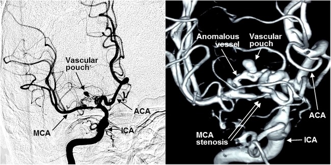
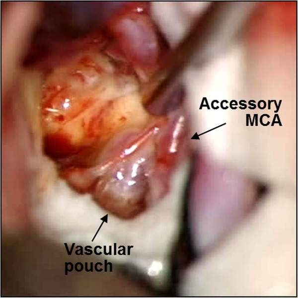
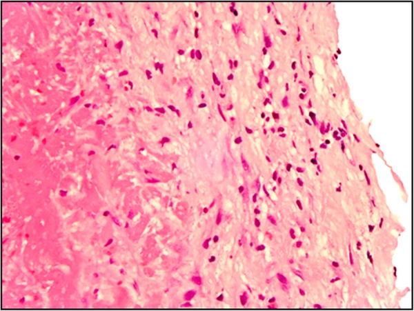
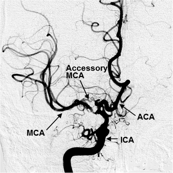
A 68-year-old man experienced a right caudate haemorrhage with intraventricular haemorrhage. Although a subarachnoid haemorrhage was not shown clearly, our investigation demonstrated an aneurysm-like vascular pouch located in the anomalous vessel arising from the A2 segment of the right anterior cerebral artery. Rupture of the vascular pouch was considered to be the cause of the caudate haemorrhage. Neck clipping was performed. In intraoperative observation, the anomalous vessel was diagnosed as a right accessory middle cerebral artery. Histopathology of the saccular wall showed only an adventitia and a fibrin layer, indicating a pseudoaneurysm. We routinely perform detailed vascular evaluation for any cerebrovascular disease. A meticulous vascular survey makes it possible to obtain valuable clues in cases such as caudate haemorrhage due to pseudoaneurysm of the accessory middle cerebral artery, leading to prevention of rebleeding.


Figures

Figure 1
Figure 1
CT scans on the first admission showing a right caudate haemorrhage with intraventricular haemorrhage and subarachnoid haemorrhage, which was not clearly demonstrated (R, right).
Figure 2
Figure 2
Preoperative right internal carotid angiograms revealing a stenosis of the M1 segment of the right MCA; the anomalous vessel originated from the A2 segment of the right ACA with an aneurysm-like vascular pouch (ICA, right internal carotid artery; ACA, right anterior cerebral artery; MCA, right middle cerebral artery).
Figure 3
Figure 3
Intraoperative photograph showing a dark red wall on part of the vascular pouch, indicating partial thrombosis (Accessory MCA, right accessory middle cerebral artery).
Figure 4
Figure 4
Histopathological photomicrograph of the saccular wall demonstrating that the arterial layer was only an adventitia and a fibrin without intima, internal elastic membrane and media. H&E stain, magnification ×400.
Figure 5
Figure 5
Postoperative right internal carotid angiogram showing patency of the right accessory MCA and exclusion of the vascular pouch (ICA, right internal carotid artery; ACA, right anterior cerebral artery; MCA, right middle cerebral artery; Accessory MCA, right accessory middle cerebral artery).

References

    1. Liliang PC, Liang CL, Lu CH et al. . Hypertensive caudate hemorrhage prognostic predictor, outcome, and role of external ventricular drainage. Stroke 2001;32:1195–200. 10.1161/01.STR.32.5.1195 - DOI - PubMed
    1. Kumral E, Evyapan D, Balkir K. Acute caudate vascular lesions. Stroke 1999;30:100–8. 10.1161/01.STR.30.1.100 - DOI - PubMed
    1. Reis CV, Zabramski JM, Safavi-Abbasi S et al. . The accessory middle cerebral artery: anatomic report. Neurosurgery 2008;63(1 Suppl 1):ONS10–13; discussion ONS13-14 10.1227/01.neu.0000335005.99756.eb - DOI - PubMed
    1. Teal JS, Rumbaugh CL, Bergeron RT et al. . Anomalies of the middle cerebral artery: accessory artery, duplication, and early bifurcation. AM J Roentgenol Radium Ther Nucl Med 1973;118:567–75. 10.2214/ajr.118.3.567 - DOI - PubMed
    1. Takahashi S, Hoshino F, Uemura K et al. . Accessory middle cerebral artery: is it a variant form of the recurrent artery of Heubner? AJNR 1989;10:563–8. - PMC - PubMed

Publication types