. 2015 Dec 30:15:114.
doi: 10.1186/s12896-015-0229-6.

Dental follicle stem cells in bone regeneration on titanium implants

Affiliations

Dental follicle stem cells in bone regeneration on titanium implants

Ondine Lucaciu et al. BMC Biotechnol. .

Abstract

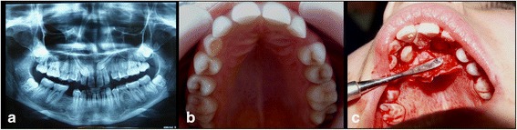
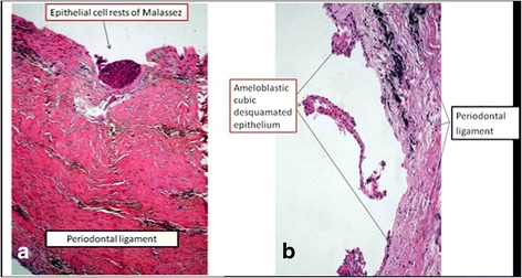
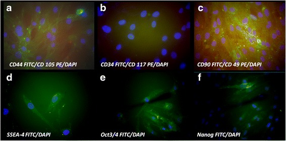
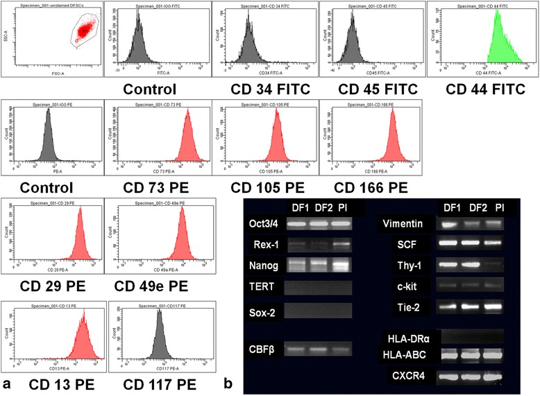
Background: We aimed to demonstrate that DF stem cells from impacted molars and canines can be used to improve bone regeneration on titanium implants surfaces. This study highlights the presence of stem cells in DF, their potential to adhere and differentiate into osteoblasts on different types of titanium surfaces.

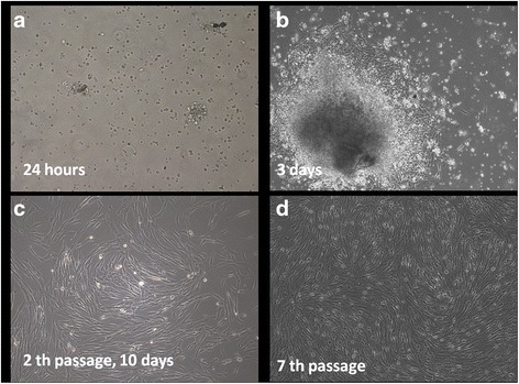
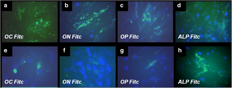
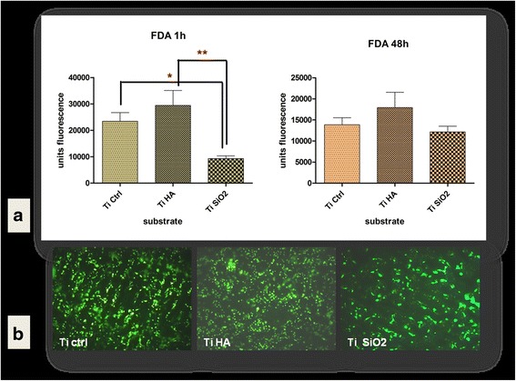
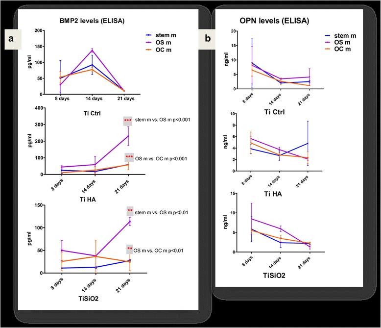
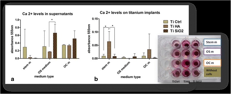
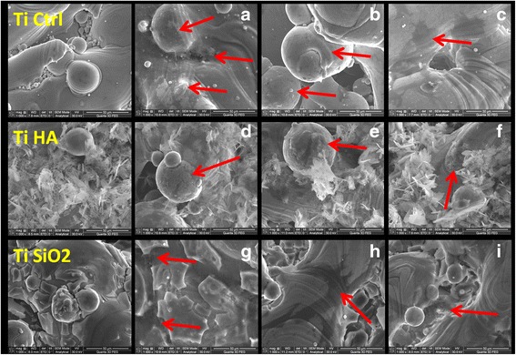
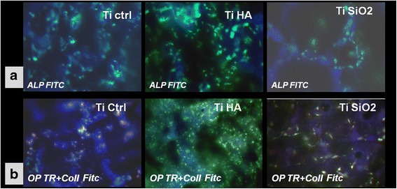
Results: Isolated cells from the harvested DF tissue from impacted canine/molars, expressed stem cells markers. Differentiation into bone cells was induced in presence or absence of BMP-2 and TGFβ1. The presence of growth factors until 28 days in medium maintained the cells in an earlier stage of differentiation with a lower level of specific bone proteins and a higher expression of alkaline phosphatase (ALP). Influence of titanium implants with different bioactive coatings, hydroxyapatite (TiHA) and with silicatitanate (TiSiO2), and porous Ti6Al7Nb implants as control (TiCtrl), was studied in terms of cell adhesion and viability. Ti HA implants proved to be more favorable for adhesion and proliferation of DF stem cells in first days of cultivation. The influence of titanium coatings and osteogenic differentiation mediums with or without growth factors were evaluated. Additional BMP-2 in the medium did not allow DF stem cells to develop a more mature phenotype, leaving them in a pre-osteogenic stage. The best sustained mineralization process evaluated by immuno-cytochemical staining, scanning electron microscopy and Ca(2+) quantification was observed for TiHA implants with a higher expression of ALP, collagen and Ca(2+) deposition. Long term culturing (70 days) on titanium surfaces of DF stem cells in standard medium without soluble osteogenic inducers, indicated that HA coating is more favorable, with the acquisition of a more mature osteoblastic phenotype as shown by immunocytochemical staining. These findings demonstrated that even in absence of exogenous osteogenic factors, TiHA implants and in a lesser extent TiCtrl and TiSiO2 implants can induce and sustain osteogenic differentiation of DF stem cells, by their chemical and topographical properties.

Conclusions: Our research demonstrated that DF stem cells have a spontaneous tendency for osteogenic differentiation and can be used for improving bone regeneration on titanium implants surfaces.


Figures

Fig. 1
Fig. 1
a radiological aspect of the two impacted canines. b clinical aspect of the two impacted canines. c surgery aspect from the removal of the dental follicle surrounding the impacted canine
Fig. 2
Fig. 2
Histological aspect of the canine dental follicle harvested tissue. a. Malassez epitelial rest and b. ameloblastic epithelia on periodontal ligament
Fig. 3
Fig. 3
Morphological aspects of isolated cells from dental follicle in contrast-phase microscopy. (a) after 24 h rounded shape cells adhere to plastic surface; (b) migrated cells after 3 days from explants presented a fibroblast-like appearance; (c) morphologically homogeneous fibroblast-like cell population was observed at 2th passage; (d) cells maintained their high proliferation rate at passage 7th (magnification ×100)
Fig. 4
Fig. 4
Immunocytochemical staining for characteristic stem cells markers, nuclei were counterstained with DAPI. (a) strong expression for CD 44 FITC and weak positivity for CD 105 PE; (b) negative staining for CD 34 FITC and CD 117 PE; (c) strong positivity for CD 90 FITC, weak expression of CD 49 PE; (d) strong positivity for early embryonic antigen SSEA-4 FITC; (e) the self-renewal embryonic proteins Oct3/4 FITC strong expression and (f) Nanog FITC positive in some cells (magnification ×400)
Fig. 5
Fig. 5
a Cell surface stem cell antigens analysed by flowcytometry: positive expression for CD44, CD73, CD105, CD29, CD49e and CD166 and lack of expression of CD34, CD45 and CD 117 indicate a mesenchymal stem cell phenotype. b RNA molecules expression in dental follicle (DF1, DF2) and placental (Pl) mesenchymal stem cells: strong expression for Oct3/4, Nanog, SCF,CXCR4, Thy-1 and Tie-2 and HLA-ABC, and absence of HLA-DR, Sox-2,c-kit, and TERT expression. Vimentin and Rex-1 have a weaker expression and was different for DF1 and DF2
Fig. 6
Fig. 6
Fluorescence images of immunocytochemical staining of DF stem cells induced for osteogenic differentiation using simple osteogenic medium in upper panel: (a) osteocalcin (OC FITC), (b) osteonectine (ON FITC), (c) osteopontin (OP FITC) and (d) alkaline phosphatase (ALP FITC). In the lower panel are illustrated the induced expression of same osteogenic markers when DF stem cells were cultivated in presence of complex osteogenic medium: (e) OC FITC, (f) ON-FITC, (g) OP-FITC and (h) ALP-FITC. Nuclei were counterstained with DAPI (Magnification ×400)
Fig. 7
Fig. 7
a DF stem cells adhesion on titanium implants after 1 h and cell viability at 48 h evaluated by fluorescein diacetate (FDA) test (area scan) (b) Fluorescence microscopy images of FDA stained DF stem cells cultivated 7 days on titanium surfaces in standard stem cells medium (Legend: TiCtrl- Ti6Al7Nb alloy porous titanium, TiHA-titanium infiltrated with hydroxyapatite, TiSiO2-titanium infiltrated with silicatitanate) (magnification ×100)
Fig. 8
Fig. 8
a BMP-2 expression in cell culture medium of DF stem cells cultivated on titanium implants in relation with differentiation medium: standard stem cell medium (blue), simple osteogenic medium (OS) (purple) and complex osteogenic medium (OC) (orange). b Osteopontin (OPN) values obtained in cell culture medium in the same conditions of DF stem cell cultures. (Legend: TiCtrl- Ti6Al7Nb alloy porous titanium, TiHA-titanium infiltrated with hydroxyapatite, TiSiO2-titanium infiltrated with silicatitanate; stem m-stem cell medium, OS m- simple osteogenic medium; OC m- complex osteogenic medium)
Fig. 9
Fig. 9
Calcium quantification by the alizarin red method. a Ca2+ levels in supernatants harversted after 21 days, from the DF stem cell cultures on Ti Ctrl, Ti HA, Ti SiO2 implants using standard stem medium, OS and OC mediums (b) alizarin red staining of Ca2+ deposition at the surface of Ti Ctrl, Ti HA, Ti SiO2 implants, in same the conditions of cultivation for 21 days. (Legend: TiCtrl- Ti6Al7Nb alloy porous titanium, TiHA-titanium infiltrated with hydroxyapatite, TiSiO2-titanium infiltrated with silicatitanate; stem m-stem cell medium, OS m- simple osteogenic medium; OC m- complex osteogenic medium)
Fig. 10
Fig. 10
SEM images of titanium implants seeded with DF stem cells. Left panel -TiCtrl, TiHA, TiSiO2 implants without cells (a) TiCtrl with stem cell medium (arrows indicate matrix deposition) (b) TiCtrl with simple osteogenic medium (arrow-cell surrounded by bone matrix) (c) TiCtrl with complex osteogenic medium (arrow-flattened cell with numerous extensions) (d) TiHA with stem cell medium (arrow-strong matrix deposition) (e) TiHA with simple osteogenic medium (arrow-cell surrounded by bone matrix) (f) TiHA with complex osteogenic medium (g) TiSiO2 with stem cell medium (arrows indicate matrix deposition inside of grooves) (h) TiSiO2 with simple osteogenic medium (arrow-large flattened cell with numerous extensions) (i) TiSiO2 with complex osteogenic medium (arrow- embedded cells in matrix) (magnification ×1000)
Fig. 11
Fig. 11
a Fluorescence images of immunocytochemical staining for alkaline phosphatase (ALP FITC) expression after 70 days of cultivation of DF stem cells on Ti implants surface (TiCtrl-porous Ti6Al7Nb titanium, TiHA-titanium infiltrated with hydroxyapatite, TiSiO2-titanium infiltrated with silicatitanate) in standard stem cells medium (magnification ×200). b Immunocytochemical staining for osteopontin (OP marked with texas red) and collagen (Coll marked with FITC) of DF stem cells cultivated in standard conditions on Ti implants surface (TiCtrl-Ti6Al7Nb porous titanium, TiHA-titanium infiltrated with hydroxyapatite, TiSiO2-titanium infiltrated with silicatitanate) (magnification ×200)

References

    1. Honda MJ, Imaizumi M, Tsuchiiya S, Morsczeck C. Dental follicle stem cells and tissue engineering. J Oral Sci. 2010;52(4):541–52. doi: 10.2334/josnusd.52.541. - DOI - PubMed
    1. Mori G, Ballini A, Carbone C, Oranger A, Brunetti G, Di Benedetto A, et al. Osteogenic differentiation of dental follicle stem cells. Int J Med Sci. 2010;9(6):480–7. doi: 10.7150/ijms.4583. - DOI - PMC - PubMed
    1. Yao S, Pan F, Prpic V, Wise GE. Differentiation of stem cells in the dental follicle. J Dent Res. 2008;87(8):767–71. doi: 10.1177/154405910808700801. - DOI - PMC - PubMed
    1. Cho MI, Garant PR. Ultrastructural evidence of directed cell migration during initial cementoblast differentiation in root formation. J Periodontal Res. 1988;23(4):268–76. doi: 10.1111/j.1600-0765.1988.tb01371.x. - DOI - PubMed
    1. Cho MI, Garant PR. Development and general structure of the periodontium. Periodontal. 2000;24:9–27. doi: 10.1034/j.1600-0757.2000.2240102.x. - DOI - PubMed

Publication types

MeSH terms