. 2016 Mar 15;310(6):L519-31.
doi: 10.1152/ajplung.00213.2015. Epub 2015 Dec 30.

MAP1LC3B overexpression protects against Hermansky-Pudlak syndrome type-1-induced defective autophagy in vitro

Affiliations

MAP1LC3B overexpression protects against Hermansky-Pudlak syndrome type-1-induced defective autophagy in vitro

Saket Ahuja et al. Am J Physiol Lung Cell Mol Physiol. .

Abstract

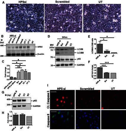
Hermansky-Pudlak syndrome (HPS) is a rare autosomal recessive disorder, and some patients with HPS develop pulmonary fibrosis, known as HPS-associated interstitial pneumonia (HPSIP). We have previously reported that HPSIP is associated with severe surfactant accumulation, lysosomal stress, and alveolar epithelial cell type II (AECII) apoptosis. Here, we hypothesized that defective autophagy might result in excessive lysosomal stress in HPSIP. Key autophagy proteins, including LC3B lipidation and p62, were increased in HPS1/2 mice lungs. Electron microscopy demonstrated a preferable binding of LC3B to the interior of lamellar bodies in the AECII of HPS1/2 mice, whereas in wild-type mice it was present on the limiting membrane in addition to the interior of the lamellar bodies. Similar observations were noted in human HPS1 lung sections. In vitro knockdown of HPS1 revealed increased LC3B lipidation and p62 accumulation, associated with an increase in proapoptotic caspases. Overexpression of LC3B decreased the HPS1 knockdown-induced p62 accumulation, whereas rapamycin treatment did not show the same effect. We conclude that loss of HPS1 protein results in impaired autophagy that is restored by exogenous LC3B and that defective autophagy might therefore play a critical role in the development and progression of HPSIP.

Keywords: Hermansky-Pudlak syndrome; Hermansky-Pudlak syndrome-associated interstitial pneumonia; alveolar epithelial cells; apoptosis; autophagy; lung fibrosis.


Figures

Fig. 1.
Fig. 1.
Defective autophagy in the alveolar epithelial type II cells (AECII) of Hermansky-Pudlak syndrome (HPS)1/2 mice. A: 3- and 9-mo-old HPS1/2, HPS1, HPS2, and wild-type (WT) mice lung homogenates were subjected to Western blotting to detect the indicated markers of autophagy. B: immunogold labeling for light chain 3β (LC3B) on lung sections of HPS1/2 and WT mice. Lamellar bodies (LBs) within AECII of WT or HPS1/2 mice lungs are depicted here. Arrow heads indicate LC3B gold labeling on the limiting membrane of LBs in WT mice. *LC3B gold labeling within the lumen of LBs in HPS1/2 mice lungs. LM, lamellar membrane of LBs. C and D: serial sections of HPS1/2 and WT mice lungs were stained for p62 and prosurfactant protein C (SP-C). Original magnification: ×200; scale bar = 100 μm (C). High-magnification images for the selected regions are depicted in D. Arrows indicate p62-positive AECII in HPS1/2 mice lung sections. Original magnification: ×400; scale bar = 50 μm (D). Representative images from 3 different experiments are shown with n = 3–5 mice. ATG, autophagy-related gene protein; TFEB, transcription factor EB; Lamp, lysosome-associated membrane protein.
Fig. 2.
Fig. 2.
Defective autophagy in AECII of lung of a patient with HPS1. Serial lung sections from 1 patient with HPS-1 and healthy donor were immunostained either for p62 or pro-SP-C. A: original magnification: ×200; scale bar = 100 μm. B: high-magnification images for the selected regions are shown, and arrows indicate p62-positive AECII in the lung sections of a patient with HPS1. Scale bar = 50 μm. C: low- and high-magnification images for negative control in which primary antibody was omitted are shown. Representative images from 3 independent experiments are shown.
Fig. 3.
Fig. 3.
siRNA-mediated knockdown of HPS1 in A549 cells results in defective autophagy and increased apoptosis. A: phase-contrast images of A549 cells after 24 h of transfection with HPS1 or scrambled siRNA or untransfected cells (UT). Scale bar = 100 μm. B: representative agarose gel image of semiquantitative PCR performed for HPS1 and β-actin from cDNA prepared from the RNA isolated from untransfected or HPS1 or scrambled or mock-transfected A549 cells. β-Actin was used as a housekeeping gene. C: densitometric quantification of HPS1 transcript levels after HPS1 knockdown compared with scrambled, mock, and untreated cells. D: representative Western blot images for LC3B, p62, and GAPDH from cell lysates prepared from untransfected, mock, or scrambled (scr) siRNA or HPS1 siRNA-transfected A549 cells after 24 h. E and F: graphical representation of densitometry analysis from Western blots shown in D is shown as ratio of LC3BII/LC3BI (E) and fold change in p62 protein level (F) compared with untransfected controls. *P < 0.05, **P < 0.01, ***P < 0.001. G: representative agarose gel image from semiquantitative RT-PCR for p62 is shown. GAPDH was used as a housekeeping gene. H: quantification of p62 transcript levels is represented as fold change compared with untransfected controls. I: representative immunofluorescence images for cleaved caspase-3 (red) and caspase-8 (green) following HPS1 or scrambled siRNA transfection or untransfected cells. DAPI (blue) was used to stain cell nuclei. Scale bar = 10 μm. Western blot images and immunofluorescence images are represented here from n = 3 independent experiments. UT1, untransfected sample 1; UT2, untransfected sample 2; NT, no template sample.
Fig. 4.
Fig. 4.
Endogenous LC3B and LAMP1 do not colocalize following HPS1 knockdown. A: A549 cells were either left untreated or were transfected with HPS1 siRNA or scrambled siRNA for 24 h followed by immunofluorescence for endogenous LC3B (red) and LAMP1 (green). B: quantification of the LC3B-LAMP1 colocalization signal is depicted as Pearson's coefficient. ***P < 0.001. C: A549 cells were transfected with GFP-LC3B plasmid for 24 h and were left untreated or transfected with HPS1 siRNA or scrambled siRNA for another 24 h followed by immunofluorescence for LAMP1. DAPI was used to stain the nuclei. Scale bar = 10 μm. D: quantification of the colocalization signals of LC3B with LAMP1 is depicted as Pearson's coefficient. ns, not significant. Representative images from n = 3 independent experiments are shown.
Fig. 5.
Fig. 5.
Overexpression of LC3B reduces HPS1 knockdown-induced p62 accumulation. A: A549 cells transfected with green fluorescent protein (GFP)-LC3B were left either untransfected or transfected with mock, HPS1, or scrambled siRNA for another 24 h. Cell lysates were subjected to Western blotting using anti-GFP antibody. Empty GFP plasmid-transfected cells were taken as controls to detect free GFP. *Bands corresponding to free GFP. B: phase-contrast images of cells indicating vacuolar structures in HPS1 siRNA transfection that are not detectable in GFP-LC3 and HPS1 siRNA-transfected cells. C–F: A549 cells were transfected with GFP-LC3B (C and D) or myc-LC3B (E and F) followed by transfection with scrambled or HPS1 siRNA for 24 h. Western blots and densitometry quantification for the indicated proteins show fold change compared with untransfected controls (D and F). G and H: representative Western blot images depicting p62 and GAPDH (G) and quantification of p62 Western blot (H) from cells transfected with HPS1 or scrambled siRNA, which were treated with 100 mM rapamycin. *P < 0.05. Representative images and analysis from n = 3 independent experiments are shown.
Fig. 6.
Fig. 6.
Proposed model for defective autophagy following HPS1 knockdown. A: under healthy conditions in which autophagy is functional, p62 is recruited into the autophagosomes, which fuse with lysosomes to form autophagolysosomes, in which p62 and other contents are degraded. B: under conditions of HPS1 knockdown in A549 cells, fusion between autophagosomes and lysosomes is impaired, thereby resulting in defective autophagy (LC3B and p62 accumulation). C: overexpressing LC3B (GFP-LC3B is shown as example) before knocking down HPS1 leads to the fusion of GFP-LC3B-labeled autophagosomes and lysosomes, resulting in functional autophagolysosomes and eventually p62 degradation.

Similar articles

Cited by

References

    1. Anderson PD, Huizing M, Claassen DA, White J, Gahl WA. Hermansky-Pudlak syndrome type 4 (HPS-4): clinical and molecular characteristics. Hum Genet 113: 10–17, 2003. - PubMed
    1. Anikster Y, Huizing M, White J, Shevchenko YO, Fitzpatrick DL, Touchman JW, Compton JG, Bale SJ, Swank RT, Gahl WA, Toro JR. Mutation of a new gene causes a unique form of Hermansky-Pudlak syndrome in a genetic isolate of central Puerto Rico. Nat Genet 28: 376–380, 2001. - PubMed
    1. Araya J, Kojima J, Takasaka N, Ito S, Fujii S, Hara H, Yanagisawa H, Kobayashi K, Tsurushige C, Kawaishi M, Kamiya N, Hirano J, Odaka M, Morikawa T, Nishimura SL, Kawabata Y, Hano H, Nakayama K, Kuwano K. Insufficient autophagy in idiopathic pulmonary fibrosis. Am J Physiol Lung Cell Mol Physiol 304: L56–L69, 2013. - PubMed
    1. Blommaart EF, Luiken JJ, Meijer AJ. Autophagic proteolysis: control and specificity. Histochem J 29: 365–385, 1997. - PubMed
    1. Boissy RE, Zhao Y, Gahl WA. Altered protein localization in melanocytes from Hermansky-Pudlak syndrome: support for the role of the HPS gene product in intracellular trafficking. Lab Invest 78: 1037–1048, 1998. - PubMed

Publication types

LinkOut - more resources