. 2015 Dec 30;90(6):3028-43.
doi: 10.1128/JVI.02936-15.

Detection of Quiescent Infections with Multiple Elephant Endotheliotropic Herpesviruses (EEHVs), Including EEHV2, EEHV3, EEHV6, and EEHV7, within Lymphoid Lung Nodules or Lung and Spleen Tissue Samples from Five Asymptomatic Adult African Elephants

Affiliations

Detection of Quiescent Infections with Multiple Elephant Endotheliotropic Herpesviruses (EEHVs), Including EEHV2, EEHV3, EEHV6, and EEHV7, within Lymphoid Lung Nodules or Lung and Spleen Tissue Samples from Five Asymptomatic Adult African Elephants

Jian-Chao Zong et al. J Virol. .

Abstract

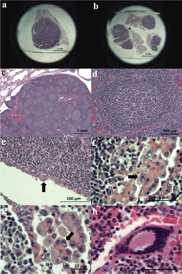
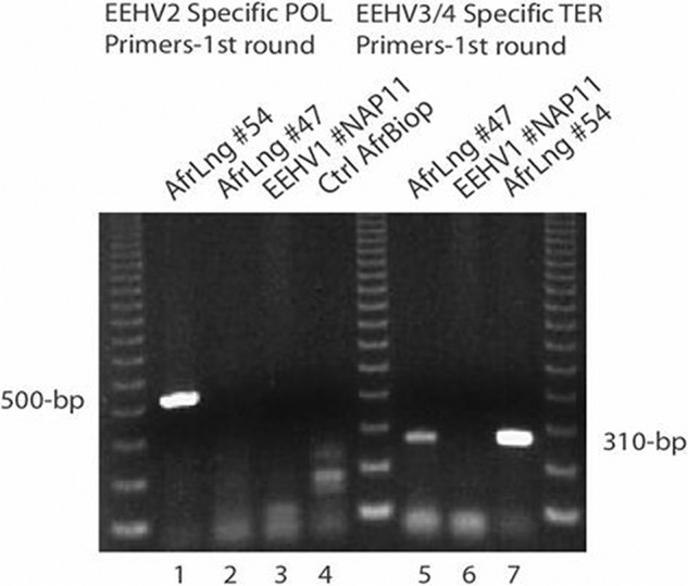
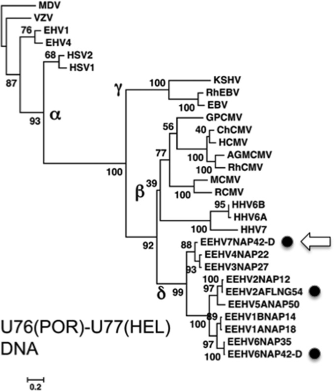
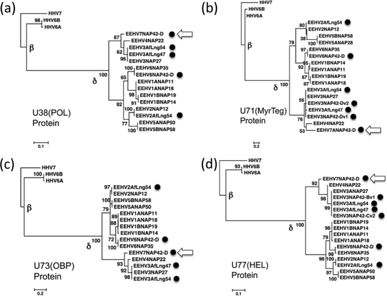
More than 80 cases of lethal hemorrhagic disease associated with elephant endotheliotropic herpesviruses (EEHVs) have been identified in young Asian elephants worldwide. Diagnostic PCR tests detected six types of EEHV in blood of elephants with acute disease, although EEHV1A is the predominant pathogenic type. Previously, the presence of herpesvirus virions within benign lung and skin nodules from healthy African elephants led to suggestions that African elephants may be the source of EEHV disease in Asian elephants. Here, we used direct PCR-based DNA sequencing to detect EEHV genomes in necropsy tissue from five healthy adult African elephants. Two large lung nodules collected from culled wild South African elephants contained high levels of either EEHV3 alone or both EEHV2 and EEHV3. Similarly, a euthanized U.S. elephant proved to harbor multiple EEHV types distributed nonuniformly across four small lung nodules, including high levels of EEHV6, lower levels of EEHV3 and EEHV2, and a new GC-rich branch type, EEHV7. Several of the same EEHV types were also detected in random lung and spleen samples from two other elephants. Sanger PCR DNA sequence data comprising 100 kb were obtained from a total of 15 different strains identified, with (except for a few hypervariable genes) the EEHV2, EEHV3, and EEHV6 strains all being closely related to known genotypes from cases of acute disease, whereas the seven loci (4.0 kb) obtained from EEHV7 averaged 18% divergence from their nearest relative, EEHV3. Overall, we conclude that these four EEHV species, but probably not EEHV1, occur commonly as quiescent infections in African elephants.

Importance: Acute hemorrhagic disease characterized by high-level viremia due to infection by members of the Proboscivirus genus threatens the future breeding success of endangered Asian elephants worldwide. Although the genomes of six EEHV types from acute cases have been partially or fully characterized, lethal disease predominantly involves a variety of strains of EEHV1, whose natural host has been unclear. Here, we carried out genotype analyses by partial PCR sequencing of necropsy tissue from five asymptomatic African elephants and identified multiple simultaneous infections by several different EEHV types, including high concentrations in lymphoid lung nodules. Overall, the results provide strong evidence that EEHV2, EEHV3, EEHV6, and EEHV7 represent natural ubiquitous infections in African elephants, whereas Asian elephants harbor EEHV1A, EEHV1B, EEHV4, and EEHV5. Although a single case of fatal cross-species infection by EEHV3 is known, the results do not support the previous concept that highly pathogenic EEHV1A crossed from African to Asian elephants in zoos.


Figures

FIG 1
FIG 1
Examples of EEHV-associated pathology in lung nodules from culled adult South African elephants. Shown are photomicrographs from hematoxylin-and-eosin-stained sections of formalin-fixed, paraffin-embedded archival blocks prepared at necropsy from healthy adult African elephants culled at Kruger National Park in 1997. (a and b) Low-power images of four examples of large lymphoid nodules in lung tissue; (c) cross-sectional image of a large lung nodule showing multiple lymphocyte-containing follicular areas; (d) more detailed higher-power image of a region with extensive follicular hyperplasia; (e) presumed viral nuclear inclusion body within an isolated epithelial cell; (f and g) higher-power images of representative clusters of enlarged alveolar epithelial cells in the area surrounding the follicular mass; (h) higher-power image of a representative syncytial cell cluster flanking the follicular mass. Arrows indicate likely nuclei with viral inclusion bodies.
FIG 2
FIG 2
Detection of both EEHV2 and EEHV3 DNAs in lung nodules from two culled adult South African elephants by PCR amplification. Shown is agarose gel electrophoretic separation of ethidium bromide-stained first-round PCR products obtained by using EEHV2-specific POL primers LGH6525 and LGH7440 (500 bp) (lanes 1 to 4) and EEHV3- and EEHV4-specific TER primers LGH6707 and LGH6708 (310 bp) (lanes 5 to 7). Lanes: 1, AfLng47; 2, AfLng54; 3, Asian elephant EEHV1A case NAP11 heart; 4, negative-control African elephant vestibular lesion biopsy specimen; 5, AfLng47; 6, Asian elephant EEHV1A case NAP11 heart; 7, AfLng54. Three size marker lanes contain multimerized 123-bp ladders.
FIG 3
FIG 3
DNA-level phylogenetic tree for African elephant lung nodule strains at the combined POR-HEL gene locus. The diagram presents linear distance-based Bayesian nucleotide dendrograms generated in MEGA5 with MUSCLE illustrating the genetic relationships at the U76(POR)-U77(HEL) PCR gene locus (628 bp) among five of the EEHV2, EEHV6, and EEHV7 strains (solid circles) from African elephant lung nodules compared to their previously described Proboscivirus orthologues EEHV1, EEHV2, EEHV3, EEHV4, EEHV5, and EEHV6 from hemorrhagic disease cases as well as selected key herpesviruses from all three other mammalian subfamilies (alpha-, beta-, and gammaherpesviruses). In comparison, the probosciviruses belong to the proposed Deltaherpesvirus subfamily (δ). The position of the novel EEHV7 version is also indicated with an arrow. Marek's disease alphaherpesvirus (MDV) was used as the outgroup. Bootstrap values (percentages from 1,000 reiterations) and a distance scale of 0.2 are shown. Alphaherpesvirinae (α) include Marek's disease virus (MDV), varicella-zoster virus (VZV), equine herpesvirus 1 (EEHV1), EEHV4, herpes simplex virus 1 (HSV-1), and HSV-2; Gammaherpesvirinae (γ) include Kaposi's sarcoma-associated herpesvirus (KSHV), Epstein-Barr virus (EBV), and rhesus EBV (RhEBV); and Betaherpesvirinae (β) include human cytomegalovirus (HCMV), chimpanzee CMV (ChCMV), RhCMV, African green monkey CMV (AgmCMV), guinea pig CMV (GPCMV), mouse CMV (MCMV), rat CMV (RCMV), and the Roseolovirus species human herpesvirus 6A (HHV-6A), HHV-6B, and HHV-7.
FIG 4
FIG 4
Protein-level phylogenetic trees comparing POL, MyrTeg, OBP, and HEL from African elephant lung nodules with all other known EEHV types. The diagrams present distance-based linear Bayesian amino acid dendrograms generated in MEGA5 with MUSCLE illustrating the genetic relationships among four proteins each from the EEHV2, EEHV3, EEHV6, and EEHV7 strains detected in African elephant lung nodules (solid circles) compared to their Proboscivirus (EEHV) orthologues from other Asian and African elephant hemorrhagic disease cases. The positions of the novel EEHV7 versions are also indicated with arrows. The Proboscivirus genus belongs to the proposed Deltaherpesviridae family (δ). The HHV-6A, HHV-6B, and HHV-7 orthologues within the Roseolovirus genus of the Betaherpesviridae (β) are the only non-Proboscivirus examples included for reference comparisons here. The HHV-7 version is used as the outgroup in all panels. Bootstrap values (percent) and distance scales of 0.1 or 0.2 are shown. (a) U38(POL), a DNA polymerase of 128 aa (18 examples). (b) U71(MyrTeg), a myristylated tegument protein of 48 aa (20 examples). (c) U73(OBP), an origin binding protein of 125 aa (18 examples). (d) U77(HEL), a helicase subunit of 163 aa (20 examples).

References

    1. Richman LK, Montali RJ, Garber RL, Kennedy MA, Lehnhardt J, Hildebrandt T, Schmitt D, Hardy D, Alcendor DJ, Hayward GS. 1999. Novel endotheliotropic herpesviruses fatal for Asian and African elephants. Science 283:1171–1176. - PubMed
    1. Davison AJ, Eberle R, Ehlers B, Hayward GS, McGeoch DJ, Minson AC, Pellett PE, Roizman B, Studdert MJ, Thiry E. 2009. The order Herpesvirales. Arch Virol 154:171–177. doi:10.1007/s00705-008-0278-4. - DOI - PMC - PubMed
    1. Latimer E, Zong J-C, Heaggans SY, Richman LK, Hayward GS. 2011. Detection and evaluation of novel herpesviruses in routine and pathological samples from Asian and African elephants: identification of two new probosciviruses (EEHV5 and EEHV6) and two new gammaherpesviruses (EGHV3B and EGHV5). Vet Microbiol 147:28–41. doi:10.1016/j.vetmic.2010.05.042. - DOI - PMC - PubMed
    1. Richman LK, Zong J-C, Latimer E, Lock J, Fleischer RC, Heaggans SY, Hayward GS. 2014. Elephant endotheliotropic herpesviruses EEHV1A, EEHV1B, and EEHV2 from cases of hemorrhagic disease are highly diverged from other mammalian herpesviruses and may form a new subfamily. J Virol 88:13523–13546. doi:10.1128/JVI.01673-14. - DOI - PMC - PubMed
    1. Zong J-C, Latimer EM, Long SY, Richman LK, Heaggans SY, Hayward GS. 2014. Comparative genome analysis of four elephant endotheliotropic herpesviruses, EEHV3, EEHV4, EEHV5, and EEHV6, from cases of hemorrhagic disease or viremia. J Virol 88:13547–13569. doi:10.1128/JVI.01675-14. - DOI - PMC - PubMed

Publication types

LinkOut - more resources