. 2015 Dec 30;90(6):2959-70.
doi: 10.1128/JVI.02507-15.

Human Cytomegalovirus US28 Is Important for Latent Infection of Hematopoietic Progenitor Cells

Affiliations

Human Cytomegalovirus US28 Is Important for Latent Infection of Hematopoietic Progenitor Cells

Monica S Humby et al. J Virol. .

Abstract

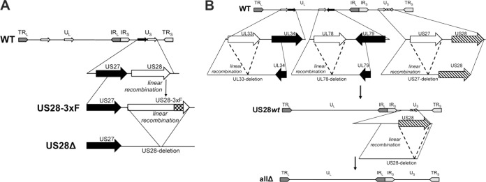
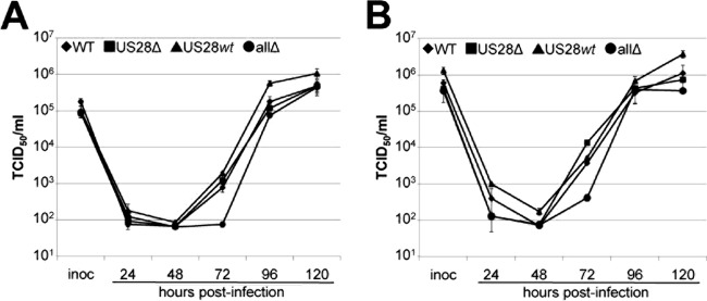
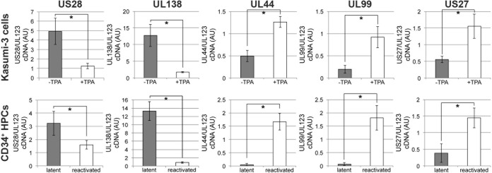
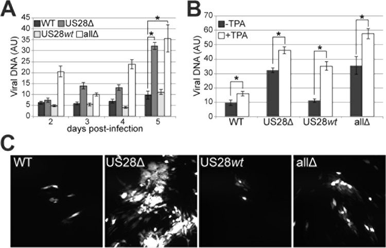
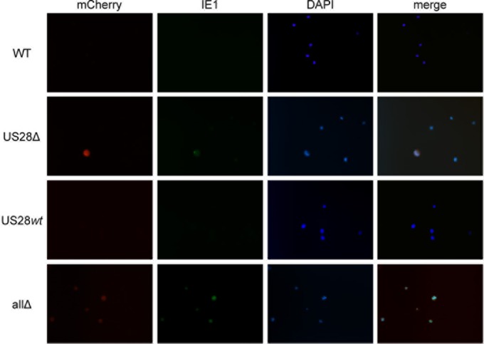
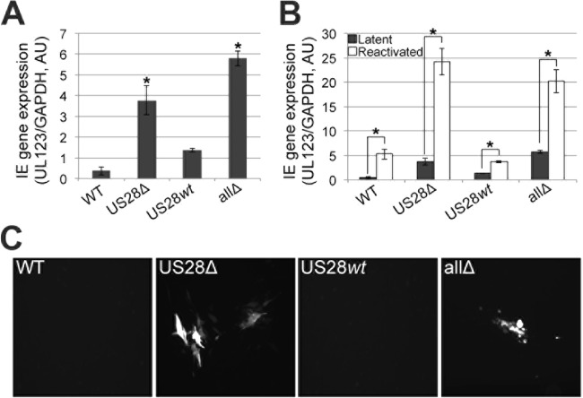
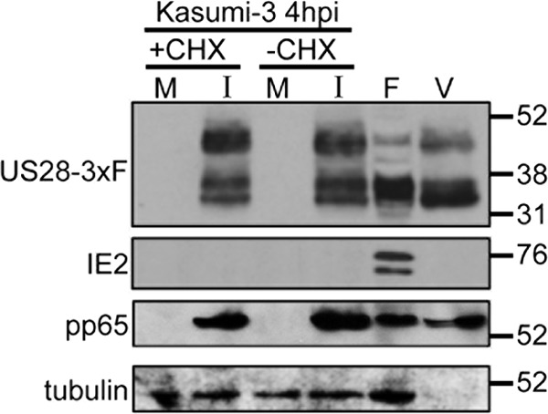
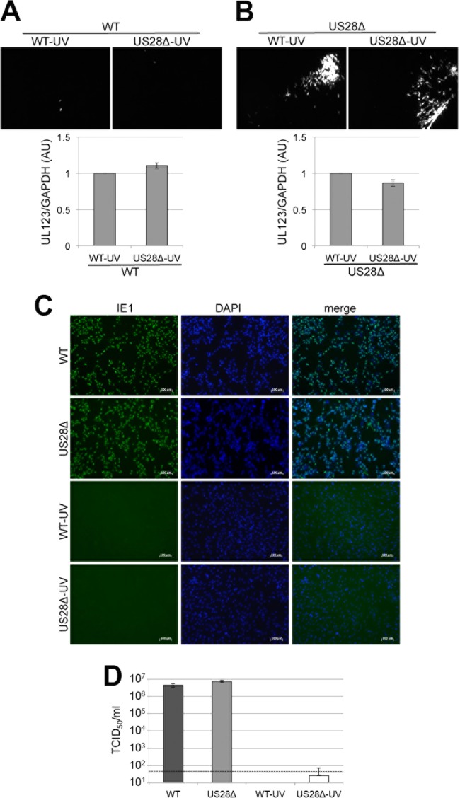
Human cytomegalovirus (HCMV) resides latently in hematopoietic progenitor cells (HPCs). During latency, only a subset of HCMV genes is transcribed, including one of the four virus-encoded G protein-coupled receptors (GPCRs), US28. Although US28 is a multifunctional lytic protein, its function during latency has remained undefined. We generated a panel of US28 recombinant viruses in the bacterial artificial chromosome (BAC)-derived clinical HCMV strain TB40/E-mCherry. We deleted the entire US28 open reading frame (ORF), deleted all four of the viral GPCR ORFs, or deleted three of the HCMV GPCRs but not the US28 wild-type protein. Using these recombinant viruses, we assessed the requirement for US28 during latency in the Kasumi-3 in vitro latency model system and in primary ex vivo-cultured CD34(+) HPCs. Our data suggest that US28 is required for latency as infection with viruses lacking the US28 ORF alone or in combination with the remaining HCMV-encoded GPCR results in transcription from the major immediate early promoter, the production of extracellular virions, and the production of infectious virus capable of infecting naive fibroblasts. The other HCMV GPCRs are not required for this phenotype as a virus expressing only US28 but not the remaining virus-encoded GPCRs is phenotypically similar to that of wild-type latent infection. Finally, we found that US28 copurifies with mature virions and is expressed in HPCs upon virus entry although its expression at the time of infection does not complement the US28 deletion latency phenotype. This work suggests that US28 protein functions to promote a latent state within hematopoietic progenitor cells.

Importance: Human cytomegalovirus (HCMV) is a widespread pathogen that, once acquired, remains with its host for life. HCMV remains latent, or quiescent, in cells of the hematopoietic compartment and upon immune challenge can reactivate to cause disease. HCMV-encoded US28 is one of several genes expressed during latency although its biological function during this phase of infection has remained undefined. Here, we show that US28 aids in promoting experimental latency in tissue culture.


Figures

FIG 1
FIG 1
Schematic of recombinant viruses. (A) BAC-derived TB40/E-mCherry was used to generate TB40/E-mCherry-US28-3×F (US28-3×F), which contains three tandem FLAG epitopes at the C terminus of the US28 ORF (54). TB40/E-mCherry-US28Δ (US28Δ) was generated by deleting the entire US28 epitope-tagged ORF (54). (B) To generate a recombinant in which US28 is the only intact HCMV GPCR ORF, we performed a series of recombination events whereby the entire coding regions for UL33, UL78, and US27 were excised and repaired by linear recombination. This resulted in a virus termed TB40/E-mCherry-US28wt (US28wt). Finally, using US28wt, we deleted US28 in its entirety, yielding TB40/E-mCherry-allΔ (allΔ) (54). TRL and TRS, long and short terminal repeat, respectively; UL and US, long and short unique region, respectively; IRL and IRS, long and short internal repeat, respectively.
FIG 2
FIG 2
US28 is dispensable for lytic grown in fibroblasts. Primary fibroblasts (NuFF-1) were infected with the viruses indicated at a multiplicity of infection of 0.5 TCID50/cell (A) or 2.0 TCID50s/cell (B). Supernatants were harvested over a time course of 120 h, and then titers for each time point were determined by TCID50 assays. Samples were analyzed in triplicate.
FIG 3
FIG 3
US28 is expressed in hematopoietic progenitor cells during latent infection. To confirm US28 expression during latency, we infected Kasumi-3 cells or primary CD34+ cells under conditions favoring a latent infection. Following 18 days in culture, each cell subset was cultured for an additional 2 days under conditions favoring either latency, using Kasumi-3 cells without TPA (23) or CD34+ cells in human long-term culture medium (51), or lytic reactivation, using Kasumi-3 cells with TPA (23) or CD34+ cells in reactivation medium (51). Cells were then harvested for RNA isolation, and the ratio of cDNA expression of US28 to UL123 was assessed by RT-qPCR in triplicate. Under latent conditions, the ratio of US28/UL123 is high, suggesting that US28 is expressed while UL123 is repressed. However, the US28/UL123 ratio nears 1 upon reactivation as both of these genes are highly expressed during lytic replication. The latent transcript UL138, the early lytic transcript UL44, the late lytic transcript UL99, and an additional HCMV GPCR, US27, are also shown as controls. *, P < 0.001. AU, arbitrary units.
FIG 4
FIG 4
US28 is important for a successful latent infection in Kasumi-3 cells. (A) Kasumi-3 cells were infected with each of the indicated viruses. For each infected cell population, extracellular DNA was collected over a 5-day time course and quantified by qPCR using primers directed at UL123. Each sample was analyzed in triplicate. *, P < 0.001 relative to the WT value at 5 days p.i. (B) Parallel cultures from the experiment shown in panel A were treated with TPA to induce lytic transcription for an additional 2 days. Extracellular viral genomes were quantified as described for panel A. *, P < 0.001. (C) To confirm that the extracellular virions produced in the experiment shown in panel A were infectious, the infected Kasumi-3 cell subsets were each cocultured with naive fibroblasts for 7 days. Viral plaques (mCherry-positive) were visualized by fluorescence microscopy.
FIG 5
FIG 5
US28Δ- and allΔ-infected Kasumi-3 cells result in IE1-positive infected cells. Kasumi-3 cells infected with each of the indicated viruses under latent conditions as described in the legend of Fig. 4 were harvested for immunofluorescence assay. Cells were stained with a monoclonal antibody directed at IE1 (clone 1B12) (56), shown in green. mCherry (red) is a marker of lytic infection; DAPI (blue) is shown as a nuclear marker. Images were collected using a 40× objective and depict representative fields for each infection.
FIG 6
FIG 6
US28Δ- and allΔ-infected primary CD34+ HPCs fail to suppress MIEP-driven transcription. (A) Cord blood-derived CD34+ cells were infected with the indicated viruses. Total RNA was isolated from each population at 5 days p.i., and IE gene expression was assessed by RT-qPCR with primers directed at UL123. Samples were normalized to cellular GAPDH levels and analyzed in triplicate. *, P < 0.001 relative to the WT level. (B) Parallel cultures from the experiment shown in panel A were treated with reactivation medium for an additional 2 days, and then RT-qPCR was performed as described for panel A. *, P < 0.001. (C) Latently infected cells from the experiment shown in panel A were cocultured with naive fibroblasts for 7 days. Fibroblast-associated viral plaques (mCherry-positive) were visualized by fluorescence microscopy.
FIG 7
FIG 7
US28 is present in extracellular virions and is brought into newly infected Kasumi-3 cells. (A) Kasumi-3 cells were left untreated (−CHX) or treated with cycloheximide (+CHX), mock infected (M) or infected with US28-3×FLAG (I), and harvested at 4 hpi. Results for cell-free, purified HCMV virions (V) and extracts from lytically infected fibroblasts (F) at 96 hpi are also depicted. US28 was detected by probing immunoblots for the FLAG epitope tag. The blot was reprobed with antibodies directed to IE2 (clone 3A9) (58), pp65 (clone 8A8) (59), and cellular tubulin as a controls. Values at the right of the blot are in kilodaltons.
FIG 8
FIG 8
Virion-associated US28 fails to complement the US28Δ latency phenotype in Kasumi-3 cells. Kasumi-3 cells were coinfected with WT and WT-UV, WT and US28Δ-UV, US28Δ and WT-UV, or US28Δ and US28Δ-UV, as indicated, at a multiplicity of infection of 1.0 PFU/cell each. Each coinfection was assessed for its ability to produce infectious virus capable of infecting naive fibroblasts by coculture (top panels), as well as for UL123 transcription (bottom panels). Fibroblast-associated plaques (mCherry-positive) were visualized by fluorescence microscopy. RT-qPCR was used to quantify UL123 transcripts relative to cellular GAPDH transcripts. For both WT and US28Δ coinfections with US28Δ-UV, UL123/GAPDH values are shown relative to those of the coinfections with WT-UV. All samples were analyzed in triplicate. Inoculum from each virus (left) was qualitatively assessed by immunofluorescence for IE1-positive nuclei using a monoclonal antibody directed at IE1 (clone 1B12) (56) (C) or quantitatively assessed by TCID50 assay (D). Samples for the experiment shown in panel D were analyzed in triplicate. The dashed line represents the level of detection.

References

    1. Britt W. 2008. Manifestations of human cytomegalovirus infection: proposed mechanisms of acute and chronic disease. Curr Top Microbiol Immunol 325:417–470. - PubMed
    1. Hakki M, Chou S. 2011. The biology of cytomegalovirus drug resistance. Curr Opin Infect Dis 24:605–611. doi:10.1097/QCO.0b013e32834cfb58. - DOI - PMC - PubMed
    1. Coronel R, Takayama S, Juwono T, Hertel L. 2015. Dynamics of human cytomegalovirus infection in CD34+ hematopoietic cells and derived Langerhans-type dendritic cells. J Virol 89:5615–5632. doi:10.1128/JVI.00305-15. - DOI - PMC - PubMed
    1. Goodrum F, Jordan CT, Terhune SS, High K, Shenk T. 2004. Differential outcomes of human cytomegalovirus infection in primitive hematopoietic cell subpopulations. Blood 104:687–695. doi:10.1182/blood-2003-12-4344. - DOI - PubMed
    1. Goodrum FD, Jordan CT, High K, Shenk T. 2002. Human cytomegalovirus gene expression during infection of primary hematopoietic progenitor cells: a model for latency. Proc Natl Acad Sci U S A 99:16255–16260. doi:10.1073/pnas.252630899. - DOI - PMC - PubMed

Publication types