. 2015 Dec 30;90(6):2740-55.
doi: 10.1128/JVI.02380-15.

Immunogenicity of a Prefusion HIV-1 Envelope Trimer in Complex with a Quaternary-Structure-Specific Antibody

Affiliations

Immunogenicity of a Prefusion HIV-1 Envelope Trimer in Complex with a Quaternary-Structure-Specific Antibody

Cheng Cheng et al. J Virol. .

Abstract

The HIV-1 envelope trimer (Env) is the target of broadly neutralizing antibodies and is being explored as a vaccine candidate to elicit protective antibodies. One of the most promising antigenic and structural mimics of HIV-1 Env is the SOSIP.664-stabilized soluble trimer from the clade A strain BG505, which is preferentially recognized by broadly neutralizing antibodies. Trimer immunization elicits high-titer neutralization of the autologous tier 2 BG505 strain; however, breadth is limited, and substantial interest has focused on understanding and improving trimer immunogenicity. We sought to improve the antigenic specificity of BG505 SOSIP.664 by reducing recognition of the variable loop 3 (V3) region, which elicits only weakly neutralizing antibodies. To stabilize the trimer in its prefusion closed conformation, we complexed trimeric BG505 SOSIP.664 with the antigen-binding fragment (Fab) of PGT145, a broadly neutralizing quaternary-structure-specific antibody. Compared to the ligand-free trimer, the PGT145 Fab-BG505 SOSIP.664 complex displayed increased melting temperature stability and reduced V3 recognition. In guinea pigs, immunization with the PGT145 Fab-BG505 SOSIP.664 complex elicited ∼100-fold lower V3-directed binding and neutralization titers than those obtained with ligand-free BG505 SOSIP.664. Both complexed and ligand-free BG505 SOSIP.664 elicited comparable neutralization of the autologous BG505 virus, and in both cases, BG505 neutralization mapped to the outer domain of gp120 for some guinea pigs. Our results indicate that it is possible to reduce immune recognition of the V3 region of the trimer while maintaining the antigenic profile needed to induce autologous neutralizing antibodies. These data suggest that appropriate modifications of trimer immunogens could further focus the immune response on key neutralization epitopes.

Importance: HIV-1 Env trimers have been proposed as preferred HIV-1 vaccine immunogens. One version, BG505 SOSIP.664, a soluble stabilized trimer, was recently shown to elicit high-titer autologous neutralizing antibodies (NAbs) in rabbits. Here we compared two immunogens: the ligand-free BG505 SOSIP.664 trimer and the same trimer bound to the antigen-binding fragment (Fab) of the PGT145 antibody, a broadly neutralizing antibody which recognizes the trimer at its membrane-distal apex. We hypothesized that the Fab-bound complex would stabilize BG505 SOSIP.664 in its prefusion closed conformation and limit reactivity to weakly neutralizing antibodies targeting the variable loop 3 (V3) region. In guinea pigs, the Fab-complexed trimer induced 100-fold lower responses to the V3 region, while both ligand-free and Fab-complexed trimers elicited similar levels of autologous NAbs. Our findings demonstrate the potential to reduce "off-target" immunogenicity while maintaining the capacity to generate autologous NAbs.


Figures

FIG 1
FIG 1
On-column purification of PGT145-trimer complex. (A) Schematic of on-column complex formation between PGT145 and the BG505 trimer. PGT145 IgG containing a protease (HRV3C) cleavage site in the hinge region was loaded onto protein A resin. The supernatant from 293F cells expressing BG505 SOSIP.664 was flowed over the protein A-PGT145 IgG column. The PGT145-trimer complex was cleaved off the column by adding HRV3C. The complex was further purified by Superdex 200 gel filtration. (B) Gel filtration profile of the eluate from panel A showing the PGT145-trimer complex and excess PGT145 Fab. (C) Gel filtration profile of the PGT145-trimer complex (in red), using complex fractions from panel B, and an overlay with the profile of the 2G12 affinity-purified BG505 trimer. Proteins were detected by the A280 (y axis), and elution volumes are shown on the x axis. (D) Coomassie blue-stained SDS-PAGE gel under reducing (R) and nonreducing (NR) conditions, showing the gel filtration-purified ligand-free Env, Fab-Env complex, and PGT145 Fab. MW, molecular weight markers.
FIG 2
FIG 2
Thermal stability of ligand-free and Fab-bound Env trimers. (A) Differential scanning fluorimetry (DSF) showed the fluorescence of Sypro orange dye as it bound to the BG505 trimer (blue), the PGT145-trimer complex (red), or PGT145 Fab (green) in response to increases in temperature and trimer unfolding. The assay was run on a thermal cycler (Bio-Rad), with the dye binding to hydrophobic pockets in response to thermally induced unfolding. (B) The melting temperature (Tm) of each sample was determined by comparison of data obtained as described for panel A from the start to the end of melting (BG505 trimer, 61 to 68.5°C; and PGT145-trimer complex, 65 to 74°C), as determined by CFX Manager software (Bio-Rad). The Tm value is displayed as the halfway point of each curve (black horizontal lines) and was determined by Boltzman sigmoidal curve fitting (black dashed lines). RFU, relative fluorescence units.
FIG 3
FIG 3
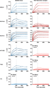
Antigenic comparison of ligand-free BG505 trimer and PGT145-trimer complex by biolayer interferometry. The binding of the ligand-free BG505 trimer (blue) and the PGT145-trimer complex (red) was assessed by capture with the following panel of antibodies: VRC01 (CD4bs directed), PGT145 (V1V2 directed), PGT151 (gp120-gp41 interface directed), 447-52D (V3 directed), F105 (CD4bs directed), and 17b (CD4-induced epitope directed). The dashed lines represent the end of the association step (300 s), and black lines represent curve fit data as calculated by Octet analysis software. When fewer than 3 curves could be fit, no fit was determined. Six concentrations from 6.25 nM to 200 nM were tested in 2-fold serial dilutions.
FIG 4
FIG 4
Antigenic comparison of ligand-free BG505 trimer and PGT145-trimer complex by MSD-ECLIA, lectin-capture ELISA, and effect of adjuvant. (A and B) Antigenicities of the ligand-free and complex trimeric proteins as measured by MSD-ECLIA. Area under the curve (AUC) values (see Materials and Methods) are shown for a panel of neutralizing (green) and nonneutralizing (red) antibodies and antibodies in the presence of CD4 (orange). (C) Antigenicities of BG505 trimer and PGT145-trimer complex as measured with a panel of V3-specific monoclonal antibodies by using lectin-capture ELISA. Monoclonal antibodies were 5-fold serially diluted starting at 10 μg/ml, and their binding to lectin-captured trimer proteins was determined. AUC values were determined with ELISA optical density values at 450 nm for different antibody dilutions. (D) Antigenicities of the ligand-free and complex trimeric proteins in the presence of poly(I·C) adjuvant as measured by MSD-ECLIA. The color style is the same as that in panel A.
FIG 5
FIG 5
Guinea pig immune responses as measured by ELISA. (A) The BG505 SOSIP.664-D7324 (D7324-tagged) trimer was captured on an ELISA plate to measure the serum response elicited by the BG505 trimer (blue) and the PGT145-trimer complex (red). Each line indicates the ELISA titers for an individual animal. Immunizations, labeled by weeks, are shown by arrows above the graph, with the first immunization performed at week 0. There was a statistically significant difference between the two immunization groups at weeks 17 and 24 (*, P < 0.05; two-tailed Mann-Whitney test). (B) V3-directed serum responses measured by V3 peptide ELISA. ELISA titers against the V3 peptide from the tier 1 MW965.26 virus are shown, as elicited by BG505 trimer (blue)- and PGT145-trimer complex (red)-immunized guinea pigs. The responses to the MW965.26 V3 peptide were lower in the PGT145-trimer complex group than in the ligand-free trimer group (*, P < 0.05; two-tailed Mann-Whitney test). (C) Anti-PGT145 Fab antibody response elicited by the BG505 trimer or the PGT145-trimer complex at prebleed (green) or week 24 (blue), as measured by ELISA. *, P < 0.05 (two-tailed Mann-Whitney test).
FIG 6
FIG 6
Immunogenicities of ligand-free BG505 trimer and PGT145-trimer complex as assessed by virus neutralization. (A) Comparison of neutralizing activities of sera from guinea pigs immunized with the BG505 trimer (blue) and the PGT145-trimer complex (red) against the autologous BG505.W6M.C2 virus (left) and the neutralization-sensitive MW965.26 virus (right). (B) Week 24 neutralization titers (geometric means with 95% confidence intervals). Animals immunized with the PGT145-trimer complex elicited an immune response with comparable autologous BG505 neutralization at week 24 (P = 0.3; two-tailed Mann-Whitney test) but reduced tier 1 virus neutralization at week 24 postvaccination (P = 0.03; two-tailed Mann-Whitney test). The response elicited with the PGT145-trimer complex was more biased toward autologous neutralizing activity, as shown by a lower ratio of the ID50 for MW965.26 to the ID50 for BG505.W6M.C2 for each animal (P = 0.03; two-tailed Mann-Whitney test). (C) Tier 1 neutralizing activity is directed to the V3 region of HIV-1 Env. (Left) Neutralization of the tier 1 virus MW965.26 correlated with anti-MW965.26 V3 peptide ELISA endpoint titers for both immunization groups at week 24. The black line represents the linear regression for all 8 animals. (Right) The MW965.26 V3 peptide was able to compete with the serum neutralization activity for MW965.26 virus in the BG505 trimer control group (P = 0.0009; two-tailed Mann-Whitney test).
FIG 7
FIG 7
Effects of elicited immune responses on Env trimer binding to CD4. Immune sera blocked Env trimer binding to CD4-expressing cells, with inhibition correlating with autologous virus neutralization activity. (A) BG505 Env trimer binding to CD4-expressing cells in the presence of various concentrations of monoclonal antibodies 447-52D (left) and VRC01 (right) in a flow cytometry-based CD4 blocking assay. (B) BG505 trimer binding to CD4-expressing cells in the presence of sera (1:50 dilution; week 24) from guinea pigs immunized with the BG505 trimer (left) and guinea pigs immunized with the PGT145-trimer complex (right) in the same flow cytometry-based CD4 blocking assay. Guinea pig numbers correspond to the names in Tables 1 and 2. (C) Serum neutralization of the autologous BG505 virus in both immunization groups correlates with the ability of sera to block CD4 binding. The values shown in the tabular part of the figure are neutralization ID50 values and percent inhibition of CD4 binding. Percent competition was calculated as the mean fluorescence on the R660 channel, with 100% competition corresponding to the fluorescence of unstained cells. The lower panel shows a graphical display of the data with a linear regression line.
FIG 8
FIG 8
Mapping of autologous BG505 virus neutralization. (A) Neutralization competition assays were performed with the indicated protein at 25 μg/ml, as described in Materials and Methods, using week 24 sera with an ID50 of >100. All competitors had mutations (CD4bs knockout mutations) to avoid attachment to cell surface CD4 in the neutralization assay. Data are shown for the sera from guinea pig 1 (GP-1), from the BG505 trimer immunization group, and GP-5, -6, and -8, from the PGT145-trimer complex immunization group. The BG505 outer domain (OD; gray) outcompeted neutralization in both GP-1 and GP-8, while eOD-GT6 (red) did not. None of the ODs competed significantly with the neutralizing activity in sera from GP-5 and GP-6. (B) Hybrid BG505 OD/eOD-GT6 construct sequences and locations on the HIV-1 gp120 (dark gray)/gp41 (light gray) protomer. The diagram gives a sequence representation of the BG505 SOSIP.664 construct and the construction of hybrid ODs. The OD contains amino acids 252 to 482, and eOD-GT6 is shown in the original amino acid sequence order of gp120. Hybrid OD mutants were made by using the BG505 backbone and eOD-GT6 sequences placed at either the C2-V3, C3-V4, or C4-V5 region, for GV3B, GV4B, or GV5B, respectively. The locations of the C2-V3 (GV3B), C3-V4 (GV4B), and C4-V5 (GV5B) regions are shown in ribbon representations of a BG505 SOSIP.664 protomer (one gp120 and one gp41), using the structure from PDB entry 4TVP. Each region inserted to make the hybrid OD is shown in red. The region that interacts with CD4 is colored yellow. (C) Neutralization competition assays as described for panel A, with hybrid OD mutants. Only GV4B OD did not compete efficiently (brown) with the neutralizing activity in immune sera from GP-1 and GP-8, suggesting that the neutralizing epitopes are proximal to the C3-V4 region.
FIG 9
FIG 9
Mapping of immune responses to the OD region of the trimer by ELISA. Binding of guinea pig IgG antibodies from week 24 immune sera to BG505 OD, eOD-GT6, or hybrid ODs was determined by ELISA. The optical densities at 450 nm are shown for different serum dilutions.

References

    1. Julien JP, Cupo A, Sok D, Stanfield RL, Lyumkis D, Deller MC, Klasse PJ, Burton DR, Sanders RW, Moore JP, Ward AB, Wilson IA. 2013. Crystal structure of a soluble cleaved HIV-1 envelope trimer. Science 342:1477–1483. doi:10.1126/science.1245625. - DOI - PMC - PubMed
    1. Pancera M, Zhou T, Druz A, Georgiev IS, Soto C, Gorman J, Huang J, Acharya P, Chuang GY, Ofek G, Stewart-Jones GB, Stuckey J, Bailer RT, Joyce MG, Louder MK, Tumba N, Yang Y, Zhang B, Cohen MS, Haynes BF, Mascola JR, Morris L, Munro JB, Blanchard SC, Mothes W, Connors M, Kwong PD. 2014. Structure and immune recognition of trimeric pre-fusion HIV-1 Env. Nature 514:455–461. doi:10.1038/nature13808. - DOI - PMC - PubMed
    1. Lyumkis D, Julien JP, de Val N, Cupo A, Potter CS, Klasse PJ, Burton DR, Sanders RW, Moore JP, Carragher B, Wilson IA, Ward AB. 2013. Cryo-EM structure of a fully glycosylated soluble cleaved HIV-1 envelope trimer. Science 342:1484–1490. doi:10.1126/science.1245627. - DOI - PMC - PubMed
    1. Wyatt R, Sodroski J. 1998. The HIV-1 envelope glycoproteins: fusogens, antigens, and immunogens. Science 280:1884–1888. doi:10.1126/science.280.5371.1884. - DOI - PubMed
    1. Ward AB, Wilson IA. 2015. Insights into the trimeric HIV-1 envelope glycoprotein structure. Trends Biochem Sci 40:101–107. doi:10.1016/j.tibs.2014.12.006. - DOI - PMC - PubMed

Publication types

MeSH terms

Substances

LinkOut - more resources