. 2016 Jan 12;113(2):E137-45.
doi: 10.1073/pnas.1517288113. Epub 2015 Dec 30.

Understanding TRPV1 activation by ligands: Insights from the binding modes of capsaicin and resiniferatoxin

Affiliations

Understanding TRPV1 activation by ligands: Insights from the binding modes of capsaicin and resiniferatoxin

Khaled Elokely et al. Proc Natl Acad Sci U S A. .

Abstract

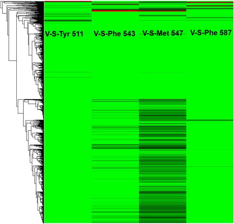
The transient receptor potential cation channel subfamily V member 1 (TRPV1) or vanilloid receptor 1 is a nonselective cation channel that is involved in the detection and transduction of nociceptive stimuli. Inflammation and nerve damage result in the up-regulation of TRPV1 transcription, and, therefore, modulators of TRPV1 channels are potentially useful in the treatment of inflammatory and neuropathic pain. Understanding the binding modes of known ligands would significantly contribute to the success of TRPV1 modulator drug design programs. The recent cryo-electron microscopy structure of TRPV1 only provides a coarse characterization of the location of capsaicin (CAPS) and resiniferatoxin (RTX). Herein, we use the information contained in the experimental electron density maps to accurately determine the binding mode of CAPS and RTX and experimentally validate the computational results by mutagenesis. On the basis of these results, we perform a detailed analysis of TRPV1-ligand interactions, characterizing the protein ligand contacts and the role of individual water molecules. Importantly, our results provide a rational explanation and suggestion of TRPV1 ligand modifications that should improve binding affinity.

Keywords: docking; heat-sensitive; ligand-gated; nociception; vanilloid.


Conflict of interest statement

The authors declare no conflict of interest.

Figures

Fig. S1.
Fig. S1.
Chemical structures of CAPS (Upper) and RTX (Lower). The three similar structural regions are described as (A) aromatic, (B) amide, ester, and (C) hydrophobic and polyring.
Fig. 1.
Fig. 1.
Molecular cavities of TRPV1. (Left) Connolly surface of the molecular cavities; the vanilloid pocket is highlighted by the black box. (Right) The cavity corresponding to the vanilloid pocket is represented as a set of space-filling spheres for the three distinct structures. The different shades of surface coloring represent distinct subpockets. Note how the shape and the volume of the pocket are significantly different in the three cases, suggesting that significant structural rearrangements of the target (induced-fit) take place on binding of the ligand.
Fig. S2.
Fig. S2.
The vanilloid pocket of the apo protein (cyan, A), the TRPV1–CAPS complex (yellow, B), and the TRPV1–RTX complex (pink, C). (A) Tyr511 is oriented downward in the apo and upward in the protein–ligand complex structures. In the apo structure, the pocket is wide in all directions, facilitating ligand to get in. Tyr511 and Arg557 are oriented in such a way allowing pocket extension. Leu573 and Leu577 are oriented away from each other, leading to a wide subpocket in this direction. This wide pocket does not support tight ligand binding. (B) In the TRPV1–CAPS complex, the pocket is smaller in size compared with the one of the apo protein due to the upward orientation of Tyr511. A small volume is observed between Met547, Asn551, and Leu515. The bulk of the cavity is close to Tyr511 to accommodate the vanillyl group of CAPS and leading to hydrophobic contacts with between with Tyr511. (C) In the TRPV1–RTX complex, the pocket volume is smaller than the one of the apo protein due to upward orientation of Tyr511. Tyr511 and Glu570 are very close in TRPV1–RTX (∼2 Å apart). A wide volume is observed between Ala665, Leu669, and Leu674 and a small volume between Met547, Asn551, and Leu515. The bulk of the cavity is close to Leu669 to accommodate the large diterpene group of RTX.
Fig. 2.
Fig. 2.
The docking poses of CAPS and RTX, determined in the TRPV1–CAPS and TRPV1–RTX complexes, respectively, and then pasted in the other two structures. (A) CAPS in the apo potein. A loose binding is observed inside the shallow binding pocket. (B) CAPS in the TRPV1–CAPS complex. (C) CAPS in TRPV1–RTX. The methoxy group of CAPS clashes with the deep pocket. (D) RTX in the apo protein. The aromatic head group of RTX clashes with the deep subpocket. (E) RTX in the TRPV1–CAPS complex. The aromatic head group and the diterpene moiety clash with the vanilloid pocket. (F) RTX in the TRPV1–RTX complex. RTX fits well inside the binding pocket.
Fig. S3.
Fig. S3.
Rotameric state of binding site residues as determined from cryoEM experiments. Shown is the experimentally determined electron density (gray shading, arbitrary isovalue) superimposed to the structure of the protein (atomic coordinates as deposited in the protein databank, gray sticks) and to our docking pose of capsaicin (orange sticks). Purple color highlights the side-chains of residues Y511, L515, F522, F543, T550, F591, I661, and A665, whose conformation is highly restrained by the density, whereas cyan indicates those (M547 and L669) for which the experimental information is less abundant.
Fig. 3.
Fig. 3.
Binding mode of CAPS based on the atomic fitting with the electron density map. (A) CAPS fit within the electron density. (B) CAPS binding mode as balls and sticks, and the surrounding amino acids as surface. (C) The docked and density-fit binding modes of CAPS are very similar and involve merely a rotation along its long axis, with little penetration of the pocket. (D) The ligand interaction model of the binding modes of CAPS. Atoms of the amino acid residues in hydrophobic contacts with the ligand are shown as spheres. Hydrogen bonds are shown as green lines. The interatomic distances between the hydrogen bond donor and acceptor are shown up to 4.0 Å.
Fig. 4.
Fig. 4.
Binding mode of RTX based on the atomic fitting with the electron density map. (A) RTX fit within the electron density. (B) RTX binding mode as balls and sticks, and the surrounding amino acids as surface. (C) The docked and density-fit binding modes of RTX are very similar. RTX is placed slightly more downward in the density-fit binding mode. (D) The ligand interaction model of the binding modes of RTX. Atoms of the amino acid residues in hydrophobic contacts with the ligand are shown as spheres. Hydrogen bonds are shown as green lines. The interatomic distances between the hydrogen bond donor and acceptor are shown up to 4.0 Å.
Fig. S4.
Fig. S4.
The interaction profile of TRPV1 modulators with the TRPV1 apo structure. Favorable interactions are shown in green and unfavorable ones in red. The docked poses (y axis) are clustered based on their contacts with the commonly interacting amino acids (x axis). (Left) Clustered interaction profiles of the ensemble of ligands. (Right) Zoom on a small cluster of six compounds, and their docking poses in the vanilloid pocket.
Fig. S5.
Fig. S5.
We are showing only the new commonly interacting amino acid residues from the interaction profile of TRPV1 modulators with TRPV1–CAPS. Favorable interactions are shown in green and unfavorable ones in red. The docked poses (y axis) are clustered based on their contacts with the commonly interacting amino acids (x axis).
Fig. 5.
Fig. 5.
Altered CAPS and RTX responses in TRPV1 mutants. TEVC oocyte electrophysiology was performed as described in Materials and Methods. (A) Representative traces, currents from repeated ramp protocols are plotted for −100, 0, and 100 mV; bottom, middle, and top traces, respectively. The effects of pH 4 (green), 1 μM CAPS (pink), and 100 μM CAPS (red) are indicated with the horizontal lines. (B) Similar representative measurements with 10 nM RTX (cyan) and 100 nM RTX (blue). (C) Summary of current amplitudes at 100 mV (mean ± SE); data were normalized to the current evoked by pH 4 at the beginning of each measurement (n = 4 for each construct both for CAPS and for RTX).
Fig. S6.
Fig. S6.
Current amplitudes induced by pH 4 in WT TRPV1 and five mutants. Data are summarized from n = 8 measurements for all channels; mean current amplitudes ± SE are plotted.
Fig. 6.
Fig. 6.
Water mapping of the TRPV1–CAPS complex. (A) Important water cluster with the putative orientation of individual water molecules. Note in particular the structural waters close to Glu-570. (B) Regions of space with large putative occupancy of uncharged water are represented in red, whereas the ones accommodating canonical waters are shown in yellow. (C) van der Waals region (green) is located close to nonpolar regions.
Fig. 7.
Fig. 7.
(A) Modification hypothesis of CAPS. Positions, which can tolerate nonpolar/polar substituents, are shown as yellow/green spheres, respectively. (B) Polar (green) and neutral (yellow) water molecules at ligand coordinate providing clues about possible ligand modification. (C) Positions permitting polar substitutions. These modifications will be stabilized by the surrounding polar amino acids. (D) Small nonpolar substitutions such as halogens supported by hydrophobic amino acid residues. (E) van der Waals substitutions supported by hydrophobic amino acid residues.
Fig. S7.
Fig. S7.
The crucial water molecules for keeping the correct geometry of the binding site of TRPV1–CAPS. The wheat surface represents the possible location of the water molecules. The two most probable water molecules are shown as ball and sticks.
Fig. S8.
Fig. S8.
Comparison between the modification plans and experimental results. A is ∼15 times more potent than CAPS by making favorable modifications in the proper positions, and B is ∼7 times less potent than CAPS due to nonrecommended modifications.
Fig. 8.
Fig. 8.
Water mapping of RTX complex. (A) Important water clusters with the putative orientation of individual water molecules. (B) Regions of space with large putative occupancy of uncharged water are represented in red, whereas the ones accommodating canonical water are shown in yellow. (C) van der Waals region is represented in green.
Fig. 9.
Fig. 9.
(A) Modification hypothesis of RTX. Positions that can tolerate nonpolar/polar substituents are shown as yellow/green spheres, respectively. (B) Polar (green) and neutral (yellow) water molecules at ligand coordinate providing clues about possible ligand modification. (C) Positions permitting polar substitutions. These modifications will be stabilized by the surrounding polar amino acids. (D) Small nonpolar substitutions such as halogens supported by hydrophobic amino acid residues. (E) van der Waals substitutions such as alkyl groups surrounded by hydrophobic amino acid residues.
Fig. S9.
Fig. S9.
Experimentally tested CAPS analogs as TRPV1 agonists. Capsaicin is shown in the upper box, illustrating the main three regions of the structure that can be modified. S1–S4 are series 1–4 of the capsaicin analogs. The three similar structural regions are described as (A) aromatic, (B) amide, ester, and (C) hydrophobic and polyring.

References

    1. Vriens J, Nilius B, Voets T. Peripheral thermosensation in mammals. Nat Rev Neurosci. 2014;15(9):573–589. - PubMed
    1. Nilius B, Owsianik G. The transient receptor potential family of ion channels. Genome Biol. 2011;12(3):218. - PMC - PubMed
    1. Clapham DE, Julius D, Montell C, Schultz G. International Union of Pharmacology. XLIX. Nomenclature and structure-function relationships of transient receptor potential channels. Pharmacol Rev. 2005;57(4):427–450. - PubMed
    1. Kunert-Keil C, Bisping F, Krüger J, Brinkmeier H. Tissue-specific expression of TRP channel genes in the mouse and its variation in three different mouse strains. BMC Genomics. 2006;7(1):159. - PMC - PubMed
    1. Ramsey IS, Delling M, Clapham DE. An introduction to TRP channels. Annu Rev Physiol. 2006;68:619–647. - PubMed

Publication types