. 2016 Mar 1;76(5):1055-1065.
doi: 10.1158/0008-5472.CAN-14-3630. Epub 2015 Dec 30.

An Immune-Inflammation Gene Expression Signature in Prostate Tumors of Smokers

Affiliations

An Immune-Inflammation Gene Expression Signature in Prostate Tumors of Smokers

Robyn L Prueitt et al. Cancer Res. .

Abstract

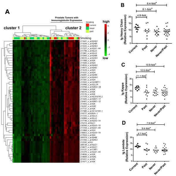
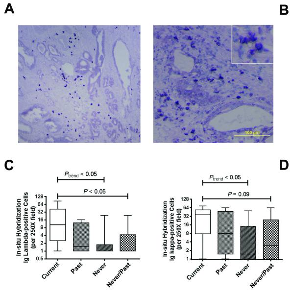
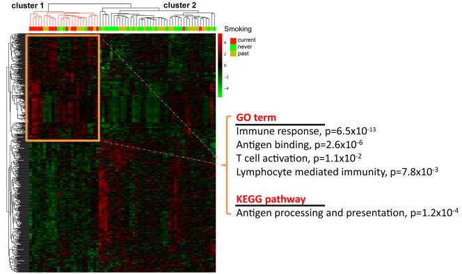
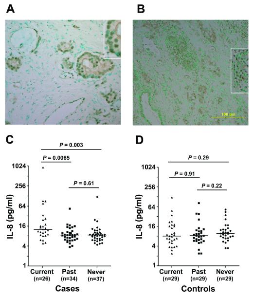
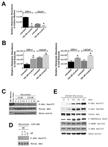
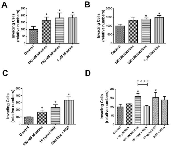
Smokers develop metastatic prostate cancer more frequently than nonsmokers, suggesting that a tobacco-derived factor is driving metastatic progression. To identify smoking-induced alterations in human prostate cancer, we analyzed gene and protein expression patterns in tumors collected from current, past, and never smokers. By this route, we elucidated a distinct pattern of molecular alterations characterized by an immune and inflammation signature in tumors from current smokers that were either attenuated or absent in past and never smokers. Specifically, this signature included elevated immunoglobulin expression by tumor-infiltrating B cells, NF-κB activation, and increased chemokine expression. In an alternate approach to characterize smoking-induced oncogenic alterations, we also explored the effects of nicotine in human prostate cancer cells and prostate cancer-prone TRAMP mice. These investigations showed that nicotine increased glutamine consumption and invasiveness of cancer cells in vitro and accelerated metastatic progression in tumor-bearing TRAMP mice. Overall, our findings suggest that nicotine is sufficient to induce a phenotype resembling the epidemiology of smoking-associated prostate cancer progression, illuminating a novel candidate driver underlying metastatic prostate cancer in current smokers.


Figures

Figure 1
Figure 1. Immunoglobulin expression in prostate tumors from current, past, and never smokers
(A) Hierarchical cluster analysis separates 47 prostate tumors (9 current, 21 past, and 17 never smokers) into 2 clusters based on immunoglobulin expression. Heatmap shows up-regulated immunoglobulin expression in cluster 2 (red color intensity). Current smokers are over-represented in this cluster (8/21 vs. 1/26 in cluster 1, P < 0.01) while the “low immunoglobulin” cluster 1 was enriched for tumors from never smokers (13/26 vs. 4/21 in cluster 2, P = 0.03). Patients’ smoking status is shown for each tumor above the heatmap. Shown are array-based expression data with corresponding probeset IDs. (B-D) Increased expression of immunoglobulin (Ig) heavy constant mu, kappa constant, and lambda locus in tumors from current smokers by qRT-PCR analysis. *Different at P < 0.05 between current smokers (n = 8) and past smokers (n = 10) or never (n = 12). qRT-PCR was performed on a subset of tumors from the microarray analysis.
Figure 2
Figure 2. Immunoglobulin expression by In situ hybridization
(A,B) Shown is the expression of kappa light chain mRNA in a representative prostate tumor from a current smoker in two different fields. In situ hybridization (ISH) depicts kappa light chain mRNA expression in lymphocytic infiltrates. The dark blue nitroblue tetrazolium chromogene reveals the predominant stromal localization of the immunoglobulin-positive B lymphocytes. Counterstain: Haematoxylin. (C,D) Analysis of ISH results. Numbers of Ig lambda- and Ig kappa-positive B lymphocytes correlated with smoking status [Spearman rank correlation test (Ptrend) for current (n = 6), past (n = 7), never (n = 9) smokers] and were increased in current smokers versus never/past smokers (Wilcoxon rank-sum test). ISH-positive lymphocytes were counted per 250x field. Shown are box plots with minimum and maximum values (whiskers) and the median as line in the box.
Figure 3
Figure 3. Up-regulation of genes in immune-related pathways is a characteristic of tumors from current smokers
Hierarchical cluster analysis with 601 probesets representing genes that were differentially expressed between current vs. past and current vs.past/never smokers. The expression pattern of these genes separates the 67 tumors (16 current, 28 past, and 23 never smokers) into 2 clusters. Cluster 1 represents tumors with up-regulated expression of immune response-related genes as shown by the Go term and KEGG pathway association. This cluster is highly enriched for tumors from current smokers (P < 0.001). Overexpressed genes in cluster 2 did not have a significant pathway or GO term association.
Figure 4
Figure 4. Nuclear accumulation of NF-κB and increased IL-8 among prostate cancer patients who smoke
(A,B) Nuclear accumulation of NF-κB P-Ser536 is shown for tumors from a past (B, 200x) and current (D, 100x) smoker (magnified in the insets). Counterstain: Methyl green. C. Cancer patients who are current smokers have increased interleukin-8 (IL-8) plasma levels when compared with past or never smokers. Median increase in current smokers was 1.5-fold. D. Current smokers without the disease did not show elevated IL-8 plasma levels. P values were calculated with the Mann-Whitney test. A Kruskal-Wallis test for differences between the three smoking groups indicated existing differences among cases (P = 0.006) but not the controls (P = 0.46).
Figure 5
Figure 5. Increased glutamine consumption and Akt activation in nicotine-treated human prostate cancer cells
22Rv1 and LNCaP cells were treated with 100 nM nicotine for 48 hours and glutamine levels were measured in culture medium and cell pellets. A. Treatment of 22Rv1 and LNCaP cells with nicotine depleted glutamine in the culture medium. B. Nicotine treatment of 22Rv1 and LNCaP cells significantly increased glutamine and glutamate levels in their cell extracts. Glutamate is the intracellular oxidation product of glutamine and was not detectable in cell culture medium. Experiments were done in triplicates. Shown is mean ± SD. * statistically different from untreated (two-sided t-test, P < 0.05). C. Nicotine at 10 nM and 100 nM concentrations induced Akt phosphorylation in 22Rv1 cells, which was inhibited by mecamylamine (MC), an antagonist of nicotinic acetylcholine receptors (D). E. Nicotine treatment also induced phosphorylation of known downstream targets of Akt signaling, such as GSK3β, human Mdm2 (HDM2), and BAD. In D, cells were exposed to nicotine −/+ MC for 2 hours.
Figure 6
Figure 6. Nicotine enhances matrigel invasion of human prostate cancer cell lines
(A) 22Rv1 and (B) PC-3 cells were were plated in invasion chambers and were treated with nicotine. (C) 22Rv1 cells were treated with nicotine, hepatocyte growth factor (HGF), or both, to assess whether nicotine and HGF have a synergistic effect on matrigel invasion. (D) Nicotinic acetylcholine receptor antagonist, mecamylamine (MCA), significantly decreased nicotine-induced increase in matrigel invasion, but not HGF-induced increase in invasion, in 22Rv1 cells. Shown are mean ± SD for n = 3. *Significantly different from control (P < 0.05).

References

    1. Jemal A, Bray F, Center MM, Ferlay J, Ward E, Forman D. Global cancer statistics. CA Cancer J.Clin. 2011;61:69–90. - PubMed
    1. Hsing AW, Chokkalingam AP. Prostate cancer epidemiology. Front Biosci. 2006;11:1388–1413. - PubMed
    1. Giovannucci E, Rimm EB, Ascherio A, Colditz GA, Spiegelman D, Stampfer MJ, Willett WC. Smoking and risk of total and fatal prostate cancer in United States health professionals. Cancer Epidemiol.Biomarkers Prev. 1999;8:277–282. - PubMed
    1. Hickey K, Do KA, Green A. Smoking and prostate cancer. Epidemiol.Rev. 2001;23:115–125. - PubMed
    1. Huncharek M, Haddock KS, Reid R, Kupelnick B. Smoking as a risk factor for prostate cancer: a meta-analysis of 24 prospective cohort studies. Am.J.Public Health. 2010;100:693–701. - PMC - PubMed

Publication types