. 2016 Feb;15(2):313-22.
doi: 10.1158/1535-7163.MCT-15-0658. Epub 2015 Dec 30.

Human Leukocyte Antigen-Presented Macrophage Migration Inhibitory Factor Is a Surface Biomarker and Potential Therapeutic Target for Ovarian Cancer

Affiliations

Human Leukocyte Antigen-Presented Macrophage Migration Inhibitory Factor Is a Surface Biomarker and Potential Therapeutic Target for Ovarian Cancer

Andrea M Patterson et al. Mol Cancer Ther. 2016 Feb.

Abstract

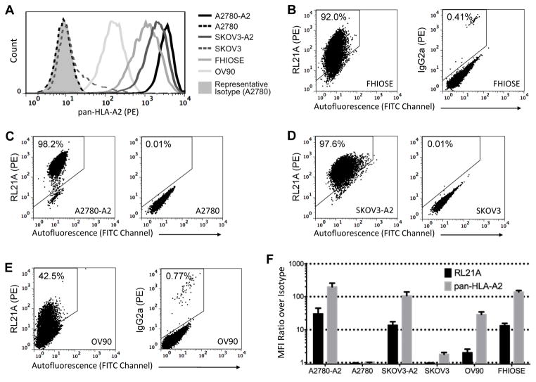
T cells recognize cancer cells via HLA/peptide complexes, and when disease overtakes these immune mechanisms, immunotherapy can exogenously target these same HLA/peptide surface markers. We previously identified an HLA-A2-presented peptide derived from macrophage migration inhibitory factor (MIF) and generated antibody RL21A against this HLA-A2/MIF complex. The objective of the current study was to assess the potential for targeting the HLA-A2/MIF complex in ovarian cancer. First, MIF peptide FLSELTQQL was eluted from the HLA-A2 of the human cancerous ovarian cell lines SKOV3, A2780, OV90, and FHIOSE118hi and detected by mass spectrometry. By flow cytometry, RL21A was shown to specifically stain these four cell lines in the context of HLA-A2. Next, partially matched HLA-A*02:01+ ovarian cancer (n = 27) and normal fallopian tube (n = 24) tissues were stained with RL21A by immunohistochemistry to assess differential HLA-A2/MIF complex expression. Ovarian tumor tissues revealed significantly increased RL21A staining compared with normal fallopian tube epithelium (P < 0.0001), with minimal staining of normal stroma and blood vessels (P < 0.0001 and P < 0.001 compared with tumor cells) suggesting a therapeutic window. We then demonstrated the anticancer activity of toxin-bound RL21A via the dose-dependent killing of ovarian cancer cells. In summary, MIF-derived peptide FLSELTQQL is HLA-A2-presented and recognized by RL21A on ovarian cancer cell lines and patient tumor tissues, and targeting of this HLA-A2/MIF complex with toxin-bound RL21A can induce ovarian cancer cell death. These results suggest that the HLA-A2/MIF complex should be further explored as a cell-surface target for ovarian cancer immunotherapy.


Conflict of interest statement

Conflict of Interest: W.H. is Co-Founder and Chief Scientist of Pure MHC LLC, a portfolio company of Emergent Technologies Inc., with which R.B. and C.M. are also affiliated. G.A., G.M., and R.D. are employees of Viventia Bio Inc.

Figures

Figure 1
Figure 1. The MIF peptide FLSELTQQL is detected in HLA-A2 from all four cancerous ovarian cell lines
(A) Overlaid extracted ion chromatograms of the 539.79 m/z ion from the synthetic MIF19-27 LC-MS run and that of the corresponding fraction from each cell line. (B) MS/MS spectrum showing the fragmentation pattern of the synthetic MIF19-27 peptide. B and Y ions are labeled along with some prominent internal fragment ions. (C) The identical fragmentation pattern to the synthesized MIF19-27 peptide in (B) was observed in all ovarian cancer cell lines at the corresponding m/z.
Figure 2
Figure 2. RL21A stains ovarian cancer cells in an HLA-A2 dependent manner
(A) Overlaid histograms comparing total HLA-A2 staining for each cell line. (B) Flow cytometry dot plots showing RL21A staining of FHIOSE compared to isotype control. (C,D) RL21A staining of HLA-A2-transfected A2780-A2 (C, left) or SKOV3-A2 (D, left) compared to the untransfected parent cells (right). (E) RL21A staining of OV90 compared to isotype control. (F) Summarized RL21A and pan-HLA-A2 mean fluorescence intensities for all lines from three independent experiments. Data are mean + SD.
Figure 3
Figure 3. RL21A tissue staining intensities
(A) Four different stains were performed on each patient tissue as indicated along the top. Three tissues are shown, each representing the indicated RL21A intensity range where x = intensity. Original magnification, x 200 (including zoomed insets). (B) Percentages of tumor and normal epithelial tissues falling within each RL21A intensity range. Actual number of tissues is indicated above each bar.
Figure 4
Figure 4. RL21A specifically targets ovarian tumor tissues
Normal FT or tumor tissues from ovarian cancer patients were stained by IHC for total HLA-A2 using BB7.2 or for the specific HLA-A2/MIF complex using RL21A. Scores integrate intensity and percent cells stained. Individual data points and median are plotted for (A) total epithelial scores, (B) matched-only epithelial scores with normal and tumor pairs connected, (C) stromal scores, and (D) blood vessel endothelial scores. Mann-Whitney U tests were performed for all but (B), which represents a Wilcoxon matched-pairs signed rank test. (E) RL21A scores only, comparing normal FT stromal and endothelial cells with the epithelial ovarian cancer cells using a Kruskal-Wallis one-way analysis of variance with Dunn’s multiple comparisons. (F) Normal FT tissue with tumor focus boxed and further magnified. Black arrows, epithelial ovarian cancer metastasis. White arrows, normal epithelium. Original magnification, x 40 (top), x 200 (bottom). *p < 0.05, **p < 0.01, ***p < 0.001, ****p < 0.0001.
Figure 5
Figure 5. Toxin-bound RL21A kills HLA-A2+ ovarian cancer cells
Cells were treated with dilutions of either bare RL21A in the presence of a constant concentration of cytotoxic secondary (A), or RL21A-deB (B) for 72 h. Live cells were detected by MTS assay. Data are mean ± SD from at least three independent experiments with at least 3 replicate wells per experiment. Data are fit with sigmoidal 4-parameter logistics curves (A2780-A2 and FHIOSE) or best-fit lines (A2780).

References

    1. American Cancer Society. Cancer Facts and Figures 2015. Atlanta: 2015.
    1. Disis ML. Mechanism of action of immunotherapy. Semin Oncol. 2014;41(Suppl 5):S3–13. - PubMed
    1. Trivedi S, Jie HB, Ferris RL. Tumor antigen-specific monoclonal antibodies and induction of T-cell immunity. Semin Oncol. 2014;41:678–84. - PMC - PubMed
    1. Joo WD, Visintin I, Mor G. Targeted cancer therapy--are the days of systemic chemotherapy numbered? Maturitas. 2013;76:308–14. - PMC - PubMed
    1. Hudis CA. Trastuzumab--mechanism of action and use in clinical practice. The New England journal of medicine. 2007;357:39–51. - PubMed

Publication types

MeSH terms