

Coincident liposarcoma, carcinoid and gastrointestinal stromal tumor complicating type 1 neurofibromatosis: Case report and literature review
- PMID: 26719605
- PMCID: PMC4674501
- DOI: 10.1016/j.jor.2014.08.010
Coincident liposarcoma, carcinoid and gastrointestinal stromal tumor complicating type 1 neurofibromatosis: Case report and literature review
Abstract
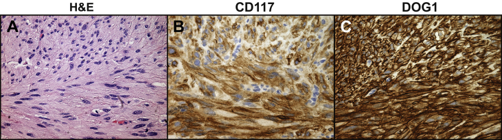
Neurofibromatosis type 1 (NF1) is associated with increased risk of multiple neoplasms. We present a case of a female patient with NF1 who presented with a rectal low-grade neuroendocrine (carcinoid) tumor. Computed tomography imaging found a well-differentiated liposarcoma and a well-circumscribed gastro-intestinal stromal tumor (GIST). Although GIST and carcinoid tumors are frequently found in NF1 patients, liposarcoma complicating NF1 is quite rare and this is the first reported case of well-differentiated liposarcoma in NF1. In summary, we report a case of coincident abdominal carcinoid tumor, GIST and well-differentiated liposarcoma, which illustrates the variability of neoplasms in NF1 patients.
Keywords: Carcinoid; GIST; Malignant peripheral nerve sheath tumor; Type 1 neurofibromatosis; Well-differentiated liposarcoma.
Figures


Similar articles
-
Rectal carcinoma and multiple gastrointestinal stromal tumors (GIST) of the small intestine in a patient with neurofibromatosis type 1: a case report.World J Surg Oncol. 2017 Aug 23;15(1):160. doi: 10.1186/s12957-017-1231-3. World J Surg Oncol. 2017. PMID: 28835241 Free PMC article.
-
Synchronous cutaneous malignant peripheral nerve sheath tumor and jejunal gastrointestinal stromal tumor and submucosal angiomyolipoma in type 1 neurofibromatosis: A case report and literature review.Medicine (Baltimore). 2023 Jan 20;102(3):e32696. doi: 10.1097/MD.0000000000032696. Medicine (Baltimore). 2023. PMID: 36701730 Free PMC article. Review.
-
Neurofibromatosis type 1, gastrointestinal stromal tumor, leiomyosarcoma and osteosarcoma: four cases of rare tumors and a review of the literature.Crit Rev Oncol Hematol. 2013 May;86(2):191-9. doi: 10.1016/j.critrevonc.2012.11.001. Epub 2012 Dec 4. Crit Rev Oncol Hematol. 2013. PMID: 23218951 Review.
-
Multiple jejunal gastrointestinal stromal tumors and Neurofibromatosis type 1: A rare association.Int J Surg Case Rep. 2021 Aug;85:106178. doi: 10.1016/j.ijscr.2021.106178. Epub 2021 Jul 7. Int J Surg Case Rep. 2021. PMID: 34274754 Free PMC article.
-
Synchronous Upper Intestinal Neurofibromas and Duodenal Periampullary Well-Differentiated Neuroendocrine Tumor Associated With Neurofibromatosis 1.Am Surg. 2021 Jan;87(1):128-130. doi: 10.1177/0003134820945236. Epub 2020 Aug 28. Am Surg. 2021. PMID: 32856931
Cited by
-
Comparison of Cancer Prevalence in Patients With Neurofibromatosis Type 1 at an Academic Cancer Center vs in the General Population From 1985 to 2020.JAMA Netw Open. 2021 Mar 1;4(3):e210945. doi: 10.1001/jamanetworkopen.2021.0945. JAMA Netw Open. 2021. PMID: 33734413 Free PMC article.
-
A Rare Case of the Coexistence of Pancreaticobiliary Maljunction and Gastrointestinal Tumor in Neurofibromatosis Type 1.Cureus. 2022 Apr 11;14(4):e24048. doi: 10.7759/cureus.24048. eCollection 2022 Apr. Cureus. 2022. PMID: 35547425 Free PMC article.
-
Spectrum of gastrointestinal lesions of neurofibromatosis type 1: a pictorial review.Insights Imaging. 2018 Oct;9(5):661-671. doi: 10.1007/s13244-018-0648-8. Epub 2018 Sep 4. Insights Imaging. 2018. PMID: 30187267 Free PMC article. Review.
-
Gastrointestinal Stromal Tumor (GIST) and Synchronous Intra-Abdominal Liposarcoma: A Report of Two Rare Cases and Literature Review.Int J Surg Oncol. 2021 Sep 1;2021:2626635. doi: 10.1155/2021/2626635. eCollection 2021. Int J Surg Oncol. 2021. PMID: 34518784 Free PMC article. Review.
-
Synchronous recurrence of concurrent colon adenocarcinoma and dedifferentiated liposarcoma.BMJ Case Rep. 2019 May 13;12(5):e228868. doi: 10.1136/bcr-2018-228868. BMJ Case Rep. 2019. PMID: 31088817 Free PMC article.
References
Publication types
LinkOut - more resources
Full Text Sources
Other Literature Sources
Research Materials
Miscellaneous
