

The healing stages of an intramedullary implanted tibia: A stress strain comparative analysis of the calcification process
- PMID: 26719629
- PMCID: PMC4674538
- DOI: 10.1016/j.jor.2015.01.016
The healing stages of an intramedullary implanted tibia: A stress strain comparative analysis of the calcification process
Abstract
Aims: The extended usage of unreamed tibial nailing resulted in reports of an increased rate of complications, especially for the distal portion of the tibia. Unreamed nailing favours biology at the expense of the achievable mechanical stability, it is therefore of interest to define the limits of the clinical indications for this method. Extra-articular fractures of the distal tibial metaphysis, meta-diaphyseal junction, and adjacent diaphysis are distinct in their management from impaction derived ''pilon'' type fractures and mid-diaphyseal fractures. The goals of this work were to gain a thorough understanding of the load-sharing mechanism between unreamed nail and bones in a fractured tibia. With this purpose a complete model of the human leg was realised, simulating a mid-diaphyseal fracture, classified as A2 type 1, according to the AO classification. The analysis of the entire chain allows to have a complete picture of the stress distribution and of the most stressed bones and soft tissues, but, more importantly can overcome problems connected with boundary conditions imposed at single bony components.
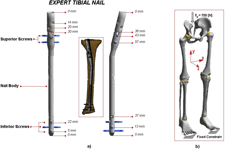
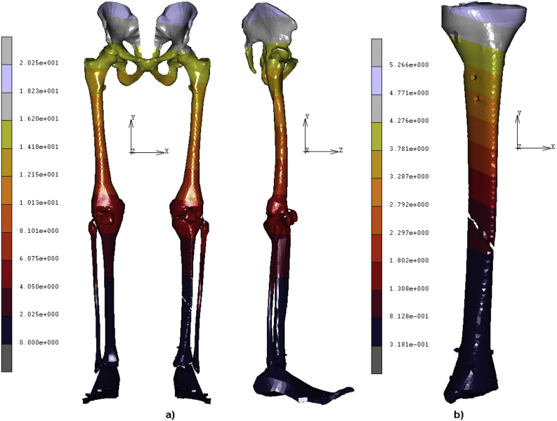
Methods: Model consists of six bony structures: pelvis, femur, patella, fibula, tibia, and a simplified lump of the feet, configured in a standing up position. Their articular cartilage layers, were simulated by 3D membranes of opportune stiffness connecting the different segments. Moreover an unreamed intra-medullary nail Expert Tibial Nail (DePuy Synthes(®)) stabilized the fractured tibia. A load of 700 N has been applied at the top of pelvis and a part the feet, at the tip, was rigidly fixed. Five different contact interfaces have been imposed at the different bony surfaces in contact.
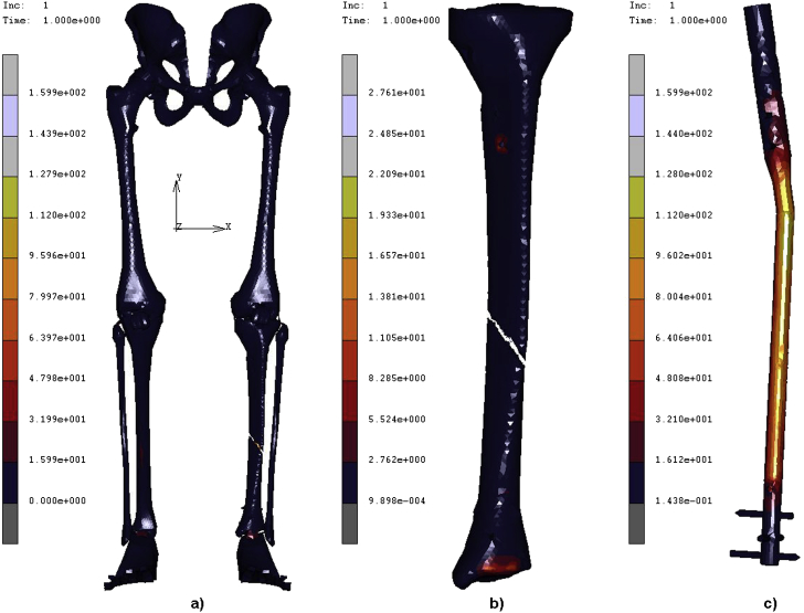
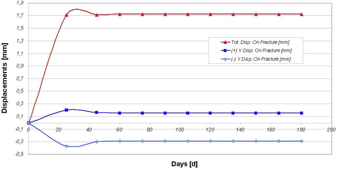
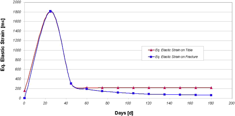
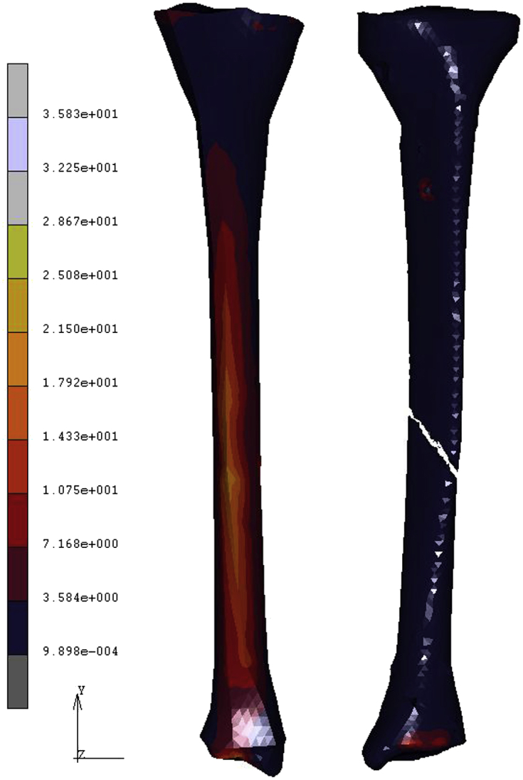
Results: Three different conditions were analysed: the initially healthy tibia, the A2 type 1 fractured tibia with the Expert tibial nail implanted, and the follow up stage after complete healing of tibia. Non-linear finite element analysis of the models were performed with Abaqus version 5.4 (Hibbitt, Karlsson and Sorensen, Inc., Pawtucket, RI) using the geometric non linearity and automatic time stepping options.
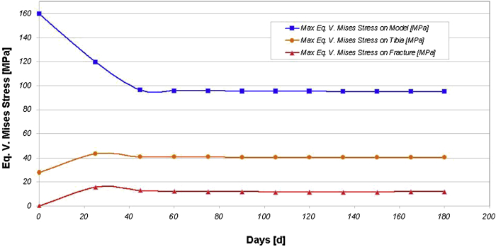
Conclusion: The obtained results reveal interesting consequences deriving by taking into account how the stress shielding can influence the integrity and resistance of bones, in order to identify the mechanical reasons for the unfavourable clinical results, and to identify borderline indications due to biomechanical factors. The evolution of treatment options for these fractures has been closely linked to developments in implant technology and surgical technique. Further developments in this area, particularly with respect to minimally invasive plating techniques and nail design are ongoing.
Keywords: Close tibial shaft fracture; Stress on tibia; Unreamed intramedullary tibial nails.
Figures
References
-
- Horne G., Iceton J., Twist J. Disability following fractures of the tibial shaft. Orthopedics. 1990;13:423–426. - PubMed
-
- Hooper G.J., Keddell R.G., Penny I.D. Conservative management or closed nailing for tibial shaft fractures. A randomised prospective trial. J Bone Joint Surg Br. 1991;73:83–85. - PubMed
-
- Haas N., Schutz M., Sudkamp N., Hoffmann R. The new unreamed AO nails for the tibia and the femur. Acta Orthop Belg. 1995;61:204–206. - PubMed
-
- Claes L., Wilke H.J., Augat P., Rubenacker S., Margevicius K.J. Effect of dynamization on gap healing of diaphyseal fractures under external fixation. Clin Biomech. 1995;10:227–234. - PubMed
-
- Burger E.H., Veldhuijzen J.P. Influence of mechanical factors on bone formations, resorption and growth in vitro. In: Hall B.K., editor. Bone. CRC Press; Boca Raton, FL: 1993. pp. 37–56.
LinkOut - more resources
Full Text Sources
Other Literature Sources
