

Disturbed spontaneous brain-activity pattern in patients with optic neuritis using amplitude of low-frequency fluctuation: a functional magnetic resonance imaging study
- PMID: 26719692
- PMCID: PMC4689287
- DOI: 10.2147/NDT.S92497
Disturbed spontaneous brain-activity pattern in patients with optic neuritis using amplitude of low-frequency fluctuation: a functional magnetic resonance imaging study
Abstract
Objective: To use the amplitude of low-frequency fluctuation (ALFF) technique to investigate the local features of spontaneous brain activity in optic neuritis (ON) and their relationship with behavioral performance.
Materials and methods: Twelve patients with ON (four male, eight female) and twelve age-, sex-, and education status-matched healthy controls (HCs) (four male, eight female) underwent resting-state functional magnetic resonance imaging (rs-fMRI) scans. The ALFF technique was used to assess local features of spontaneous brain activity. Correlation analysis was used to explore the relationship between the observed mean ALFF values of the different areas and visual evoked potentials (VEPs) in patients with ON.
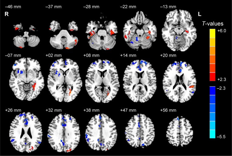
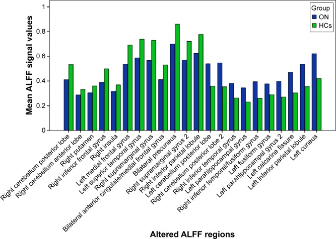
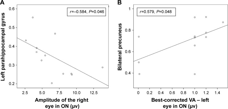
Results: Compared with HCs, patients with ON had significantly decreased ALFF values in the posterior and anterior lobes of the right cerebellum, right putamen, right inferior frontal gyrus, right insula, right supramarginal gyrus, right inferior parietal lobule, left medial frontal gyrus, left superior temporal gyrus, bilateral anterior cingulate/medial frontal gyrus, and bilateral precuneus, and significantly increased ALFF values in the posterior lobes of the left and right cerebellum, right inferior temporal gyrus, right inferior temporal/fusiform gyrus, left parahippocampal gyrus, left fusiform gyrus, left calcarine fissure, left inferior parietal lobule, and left cuneus. We found negative correlations between the mean ALFF signal value of the left parahippocampal gyrus and the VEP amplitude of the right eye in ON (r=-0.584, P=0.046), and a positive correlation between the mean ALFF signal value of the bilateral precuneus and the best-corrected visual acuity of the left eye (r=0.579, P=0.048) in patients with ON.
Conclusion: ON mainly seems to involve dysfunction in the default-mode network, cerebellum, and limbic system, which may reflect the underlying pathologic mechanism of ON.
Keywords: ALFF; fMRI; optic neuritis; resting state; spontaneous activity; visual evoked potential.
Figures
Similar articles
-
Altered intrinsic regional spontaneous brain activity in patients with optic neuritis: a resting-state functional magnetic resonance imaging study.Neuropsychiatr Dis Treat. 2015 Dec 11;11:3065-73. doi: 10.2147/NDT.S92968. eCollection 2015. Neuropsychiatr Dis Treat. 2015. PMID: 26715848 Free PMC article.
-
Altered spontaneous brain activity patterns in patients with retinal vein occlusion indicated by the amplitude of low-frequency fluctuation: A functional magnetic resonance imaging study.Exp Ther Med. 2019 Sep;18(3):2063-2071. doi: 10.3892/etm.2019.7770. Epub 2019 Jul 12. Exp Ther Med. 2019. PMID: 31410162 Free PMC article.
-
[Study on the changes of spontaneous brain activity in maintenance hemodialysis patients with end-stage renal disease based on three different resting state-functional magnetic resonance low-frequency amplitude algorithms].Zhonghua Yi Xue Za Zhi. 2021 Jan 26;101(4):265-270. doi: 10.3760/cma.j.cn112137-20200513-01524. Zhonghua Yi Xue Za Zhi. 2021. PMID: 33486935 Chinese.
-
Common and distinct patterns of intrinsic brain activity alterations in major depression and bipolar disorder: voxel-based meta-analysis.Transl Psychiatry. 2020 Oct 19;10(1):353. doi: 10.1038/s41398-020-01036-5. Transl Psychiatry. 2020. PMID: 33077728 Free PMC article. Review.
-
Altered Patterns of Amplitude of Low-Frequency Fluctuations and Fractional Amplitude of Low-Frequency Fluctuations Between Amnestic and Vascular Mild Cognitive Impairment: An ALE-Based Comparative Meta-Analysis.Front Aging Neurosci. 2021 Aug 31;13:711023. doi: 10.3389/fnagi.2021.711023. eCollection 2021. Front Aging Neurosci. 2021. PMID: 34531735 Free PMC article.
Cited by
-
The predictive potential of altered spontaneous brain activity patterns in diabetic retinopathy and nephropathy.EPMA J. 2019 Jul 5;10(3):249-259. doi: 10.1007/s13167-019-00171-4. eCollection 2019 Sep. EPMA J. 2019. PMID: 31462942 Free PMC article.
-
Voxel-Mirrored Homotopic Connectivity Is Altered in Meibomian Gland Dysfunction Patients That Are Morbidly Obese.Brain Sci. 2022 Aug 15;12(8):1078. doi: 10.3390/brainsci12081078. Brain Sci. 2022. PMID: 36009141 Free PMC article.
-
Update on central factors in myopia development beyond intraocular mechanisms.Front Neurol. 2024 Nov 18;15:1486139. doi: 10.3389/fneur.2024.1486139. eCollection 2024. Front Neurol. 2024. PMID: 39624669 Free PMC article. Review.
-
Amplitude of Low-Frequency Fluctuation to Determine Disturbed Spontaneous Brain-Activity Pattern in Patients with Diabetic Optic Neuropathy.Diabetes Metab Syndr Obes. 2023 Sep 20;16:2899-2909. doi: 10.2147/DMSO.S423111. eCollection 2023. Diabetes Metab Syndr Obes. 2023. PMID: 37753481 Free PMC article.
-
Altered spontaneous brain activity patterns in patients with hyperthyroidism exophthalmos using amplitude of low-frequency fluctuation: a resting-state fMRI study.Int J Ophthalmol. 2021 Dec 18;14(12):1957-1962. doi: 10.18240/ijo.2021.12.22. eCollection 2021. Int J Ophthalmol. 2021. PMID: 34926214 Free PMC article.
References
-
- Costello F, Coupland S, Hodge W, et al. Quantifying axonal loss after optic neuritis with optical coherence tomography. Ann Neurol. 2006;59(6):963–969. - PubMed
-
- Trip SA, Schlottmann PG, Jones SJ, et al. Retinal fiber layer axonal loss and visual dysfunction in optic nerve neuritis. An Neurol. 2005;58(3):383–391. - PubMed
LinkOut - more resources
Full Text Sources
Miscellaneous
