

GIT1 is a novel prognostic biomarker and facilitates tumor progression via activating ERK/MMP9 signaling in hepatocellular carcinoma
- PMID: 26719701
- PMCID: PMC4689273
- DOI: 10.2147/OTT.S96715
GIT1 is a novel prognostic biomarker and facilitates tumor progression via activating ERK/MMP9 signaling in hepatocellular carcinoma
Abstract
Aim: Multiple studies have revealed that G-protein-coupled receptor kinase-interacting protein 1 (GIT1) is overexpressed in many cancers and facilitates tumor progression. However, the role of GIT1 in hepatocellular carcinoma (HCC) remains unclear.
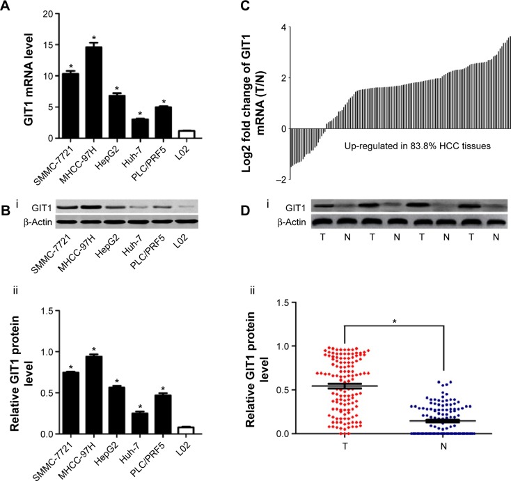
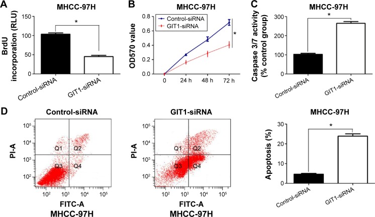
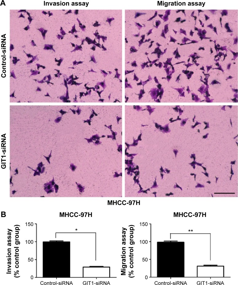
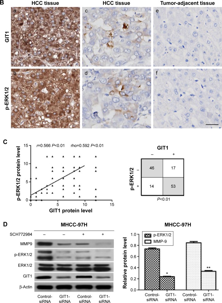
Methods: GIT1 expression was detected in cell lines and 130 pairs of HCC and matched adjacent noncancerous samples. Transwell assay, flow cytometry, caspase 3/7 activity assay, 5-bromodeoxyuridine cell proliferation assay, and 3-(4,5-dimethylthiazol-2-yl)-2,5-diphenyltetrazolium bromide assay were used to assess invasion, migration, apoptosis, and proliferation of HCC cells. Furthermore, GIT1 expression was detected by immunohistochemistry to evaluate its correlation with phospho-extracellular signal-regulated kinase (p-ERK)1/2. The regulatory effect of GIT1 on ERK1/2, p-ERK1/2, and matrix metalloproteinase-9 (MMP9) in HCC cells was confirmed by immunoblotting.
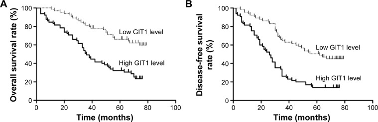
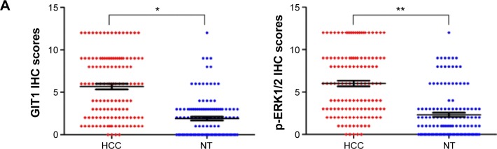
Results: In this study, we demonstrated that GIT1 was more highly expressed in HCC samples than that in non-HCC samples, and overexpression of GIT1 was correlated with clinicopathological features of poor prognosis. Clinical analysis demonstrated that GIT1 is an independent prognostic biomarker for predicting overall survival and disease-free survival of patients with HCC. In vitro studies showed that downregulation of GIT1 facilitated HCC cell apoptosis and repressed HCC cell invasion, migration, and proliferation. Overexpression of GIT1 is associated with p-ERK1/2 amplification in HCC tissues. Moreover, downregulation of GIT1 resulted in inactivation of ERK signaling and downregulation of MMP9.
Conclusion: Our findings indicate that GIT1 is an independent prognostic biomarker and facilitates HCC progression via activating ERK/MMP9 signaling.
Keywords: ERK signaling; GIT1; HCC; prognosis.
Figures
Similar articles
-
ACK1 promotes hepatocellular carcinoma progression via downregulating WWOX and activating AKT signaling.Int J Oncol. 2015 May;46(5):2057-66. doi: 10.3892/ijo.2015.2910. Epub 2015 Feb 27. Int J Oncol. 2015. PMID: 25738261
-
DDR2 facilitates hepatocellular carcinoma invasion and metastasis via activating ERK signaling and stabilizing SNAIL1.J Exp Clin Cancer Res. 2015 Sep 11;34(1):101. doi: 10.1186/s13046-015-0218-6. J Exp Clin Cancer Res. 2015. PMID: 26362312 Free PMC article.
-
GIT1 overexpression promotes epithelial-mesenchymal transition and predicts poor prognosis in hepatocellular carcinoma.Bioengineered. 2021 Dec;12(1):30-43. doi: 10.1080/21655979.2020.1855914. Bioengineered. 2021. PMID: 33258389 Free PMC article.
-
[Study on the correlation between high expression of GIT1 and M2 macrophage infiltration and prognosis in hepatocellular carcinoma].Zhonghua Gan Zang Bing Za Zhi. 2025 Mar 20;33(3):237-247. doi: 10.3760/cma.j.cn501113-20240827-00396. Zhonghua Gan Zang Bing Za Zhi. 2025. PMID: 40274549 Chinese.
-
Downregulation of castor zinc finger 1 predicts poor prognosis and facilitates hepatocellular carcinoma progression via MAPK/ERK signaling.J Exp Clin Cancer Res. 2018 Mar 5;37(1):45. doi: 10.1186/s13046-018-0720-8. J Exp Clin Cancer Res. 2018. PMID: 29506567 Free PMC article.
Cited by
-
Quercetin Inhibits the Migration and Invasion of HCCLM3 Cells by Suppressing the Expression of p-Akt1, Matrix Metalloproteinase (MMP) MMP-2, and MMP-9.Med Sci Monit. 2018 Apr 27;24:2583-2589. doi: 10.12659/MSM.906172. Med Sci Monit. 2018. PMID: 29701200 Free PMC article.
-
Snail Upregulates Transcription of FN, LEF, COX2, and COL1A1 in Hepatocellular Carcinoma: A General Model Established for Snail to Transactivate Mesenchymal Genes.Cells. 2021 Aug 26;10(9):2202. doi: 10.3390/cells10092202. Cells. 2021. PMID: 34571852 Free PMC article.
-
The Arf-GAP and protein scaffold Cat1/Git1 as a multifaceted regulator of cancer progression.Small GTPases. 2020 Mar;11(2):77-85. doi: 10.1080/21541248.2017.1362496. Epub 2017 Dec 31. Small GTPases. 2020. PMID: 28981399 Free PMC article. Review.
-
Snail collaborates with EGR-1 and SP-1 to directly activate transcription of MMP 9 and ZEB1.Sci Rep. 2017 Dec 19;7(1):17753. doi: 10.1038/s41598-017-18101-7. Sci Rep. 2017. PMID: 29259250 Free PMC article.
-
FOXP3 promotes colorectal carcinoma liver metastases by evaluating MMP9 expression via regulating S-adenosylmethionine metabolism.Ann Transl Med. 2020 May;8(9):592. doi: 10.21037/atm-20-3287. Ann Transl Med. 2020. PMID: 32566619 Free PMC article.
References
-
- Jemal A, Bray F, Center MM, Ferlay J, Ward E, Forman D. Global cancer statistics. CA Cancer J Clin. 2011;61(2):69–90. - PubMed
-
- Lafaro KJ, Demirjian AN, Pawlik TM. Epidemiology of hepatocellular carcinoma. Surg Oncol Clin N Am. 2015;24(1):1–17. - PubMed
-
- Bosetti C, Levi F, Boffetta P, Lucchini F, Negri E, La Vecchia C. Trends in mortality from hepatocellular carcinoma in Europe, 1980–2004. Hepatology. 2008;48(1):137–145. - PubMed
-
- Bosetti C, Turati F, La Vecchia C. Hepatocellular carcinoma epidemiology. Best Pract Res Clin Gastroenterol. 2014;28(5):753–770. - PubMed
LinkOut - more resources
Full Text Sources
Research Materials
Miscellaneous
