

Molecular and cytological analyses reveal distinct transformations of intestinal epithelial cells during Xenopus metamorphosis
- PMID: 26719790
- PMCID: PMC4696227
- DOI: 10.1186/s13578-015-0065-3
Molecular and cytological analyses reveal distinct transformations of intestinal epithelial cells during Xenopus metamorphosis
Abstract
Background: The thyroid hormone (T3)-induced formation of adult intestine during amphibian metamorphosis resembles the maturation of the mammalian intestine during postembryonic development, the period around birth when plasma T3 level peaks. This process involves de novo formation of adult intestinal stem cells as well as the removal of the larval epithelial cells through apoptosis. Earlier studies have revealed a number of cytological and molecular markers for the epithelial cells undergoing different changes during metamorphosis. However, the lack of established double labeling has made it difficult to ascertain the identities of the metamorphosing epithelial cells.
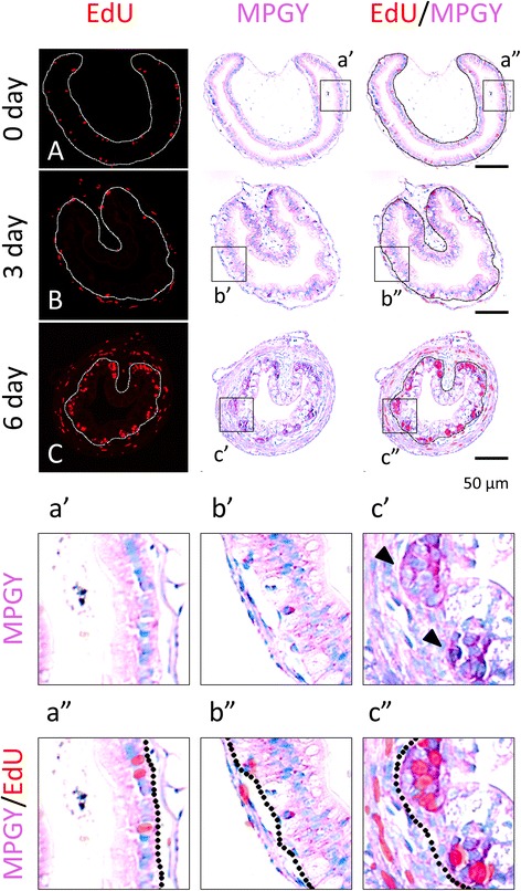
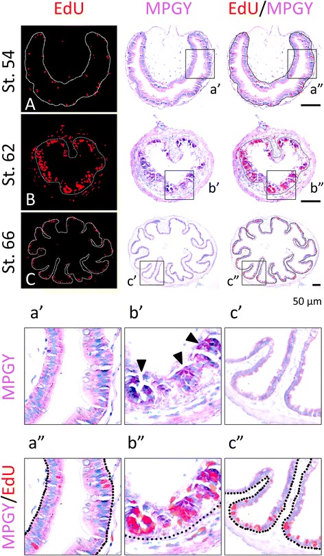
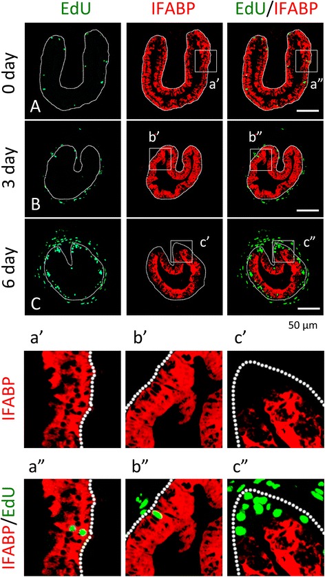
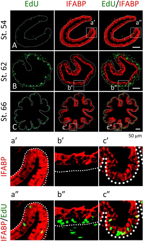
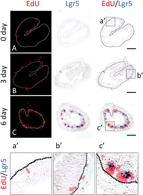
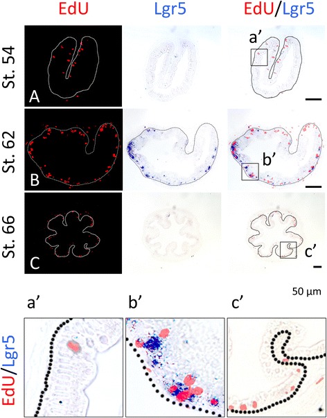
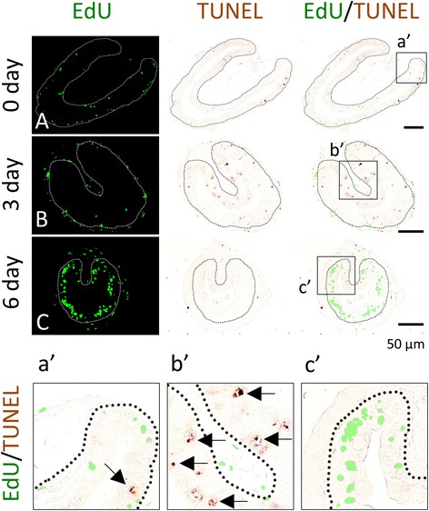
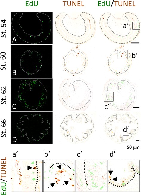
Results: Here, we carried out different double-staining with a number of cytological and molecular markers during T3-induced and natural metamorphosis in Xenopus laevis. Our studies demonstrated conclusively that the clusters of proliferating cells in the epithelium at the climax of metamorphosis are undifferentiated epithelial cells and express the well-known adult intestinal stem cell marker gene Lgr5. We further show that the adult stem cells and apoptotic larval epithelial cells are distinct epithelial cells during metamorphosis.
Conclusions: Our findings suggest that morphologically identical larval epithelial cells choose two alternative paths: programmed cell death or dedifferentiation to form adult stem cells, in response to T3 during metamorphosis with apoptosis occurring prior to the formation of the proliferating adult stem cell clusters (islets).
Keywords: Intestine; Metamorphosis; Stem cells; Thyroid hormone; Thyroid hormone receptor; Xenopus laevis.
Figures
Similar articles
-
Cell cycle activation in thyroid hormone-induced apoptosis and stem cell development during Xenopus intestinal metamorphosis.Front Endocrinol (Lausanne). 2023 May 17;14:1184013. doi: 10.3389/fendo.2023.1184013. eCollection 2023. Front Endocrinol (Lausanne). 2023. PMID: 37265708 Free PMC article. Review.
-
Spatio-temporal expression profile of stem cell-associated gene LGR5 in the intestine during thyroid hormone-dependent metamorphosis in Xenopus laevis.PLoS One. 2010 Oct 22;5(10):e13605. doi: 10.1371/journal.pone.0013605. PLoS One. 2010. PMID: 21042589 Free PMC article.
-
Analysis of Thyroid Hormone Receptor α-Knockout Tadpoles Reveals That the Activation of Cell Cycle Program Is Involved in Thyroid Hormone-Induced Larval Epithelial Cell Death and Adult Intestinal Stem Cell Development During Xenopus tropicalis Metamorphosis.Thyroid. 2021 Jan;31(1):128-142. doi: 10.1089/thy.2020.0022. Epub 2020 Jul 1. Thyroid. 2021. PMID: 32515287 Free PMC article.
-
Direct Activation of Amidohydrolase Domain-Containing 1 Gene by Thyroid Hormone Implicates a Role in the Formation of Adult Intestinal Stem Cells During Xenopus Metamorphosis.Endocrinology. 2015 Sep;156(9):3381-93. doi: 10.1210/en.2015-1190. Epub 2015 Jun 18. Endocrinology. 2015. PMID: 26086244 Free PMC article.
-
Thyroid hormone regulation of adult intestinal stem cell development: mechanisms and evolutionary conservations.Int J Biol Sci. 2012;8(8):1217-24. doi: 10.7150/ijbs.5109. Epub 2012 Oct 23. Int J Biol Sci. 2012. PMID: 23136549 Free PMC article. Review.
Cited by
-
Thyroid Hormone Receptor Is Essential for Larval Epithelial Apoptosis and Adult Epithelial Stem Cell Development but Not Adult Intestinal Morphogenesis during Xenopus tropicalis Metamorphosis.Cells. 2021 Mar 3;10(3):536. doi: 10.3390/cells10030536. Cells. 2021. PMID: 33802526 Free PMC article.
-
Thyroid hormone receptor α controls larval intestinal epithelial cell death by regulating the CDK1 pathway.Commun Biol. 2022 Feb 7;5(1):112. doi: 10.1038/s42003-022-03061-0. Commun Biol. 2022. PMID: 35132135 Free PMC article.
-
Protein arginine methyltransferase 1 regulates cell proliferation and differentiation in adult mouse adult intestine.Cell Biosci. 2021 Jun 22;11(1):113. doi: 10.1186/s13578-021-00627-z. Cell Biosci. 2021. PMID: 34158114 Free PMC article.
-
EVI and MDS/EVI are required for adult intestinal stem cell formation during postembryonic vertebrate development.FASEB J. 2018 Jan;32(1):431-439. doi: 10.1096/fj.201700424R. Epub 2017 Sep 19. FASEB J. 2018. PMID: 28928245 Free PMC article.
-
Direct activation of tRNA methyltransferase-like 1 (Mettl1) gene by thyroid hormone receptor implicates a role in adult intestinal stem cell development and proliferation during Xenopus tropicalis metamorphosis.Cell Biosci. 2020 May 4;10:60. doi: 10.1186/s13578-020-00423-1. eCollection 2020. Cell Biosci. 2020. PMID: 32391142 Free PMC article.
References
-
- Ishizuya-Oka A, Shi YB. Regulation of adult intestinal epithelial stem cell development by thyroid hormone during Xenopus laevis metamorphosis. Dev Dyn. 2007. - PubMed
-
- Muncan V, Heijmans J, Krasinski SD, Buller NV, Wildenberg ME, Meisner S, Radonjic M, Stapleton KA, Lamers WH, Biemond I, van den Bergh Weerman MA, O’Carroll D, Hardwick JC, Hommes DW, van den Brink GR. Blimp1 regulates the transition of neonatal to adult intestinal epithelium. Nat Commun. 2011;2:452. - PMC - PubMed
LinkOut - more resources
Full Text Sources
Other Literature Sources
