Comparative Study
. 2016 Jan;37(1):115-25.
doi: 10.3892/ijmm.2015.2413. Epub 2015 Nov 19.

Comparison of molecular profiles of human mesenchymal stem cells derived from bone marrow, umbilical cord blood, placenta and adipose tissue

Affiliations
Comparative Study

Comparison of molecular profiles of human mesenchymal stem cells derived from bone marrow, umbilical cord blood, placenta and adipose tissue

June Seok Heo et al. Int J Mol Med. 2016 Jan.

Abstract

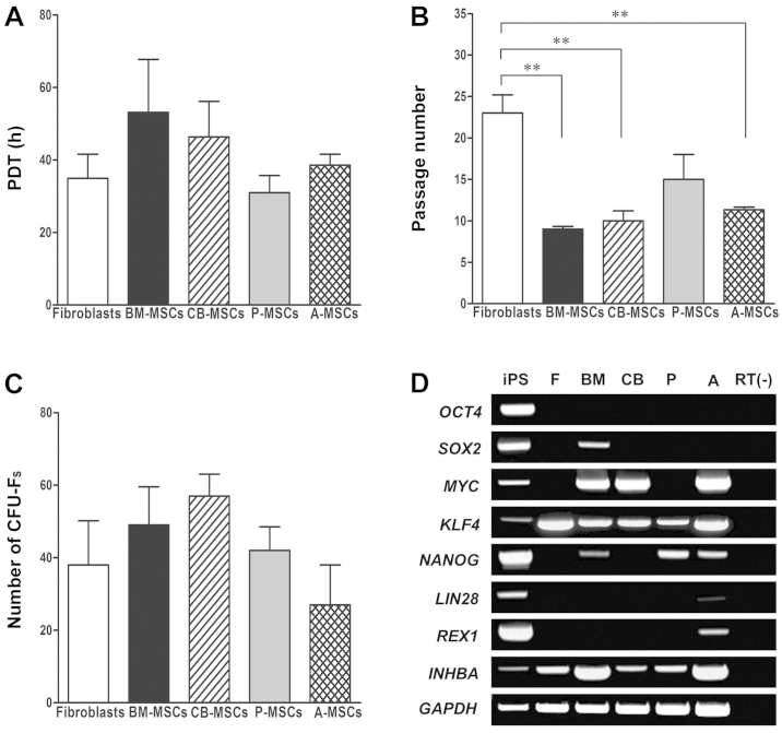
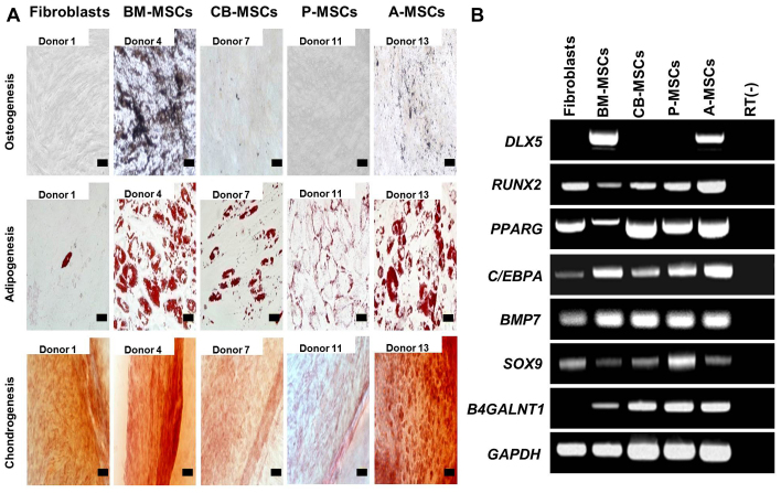
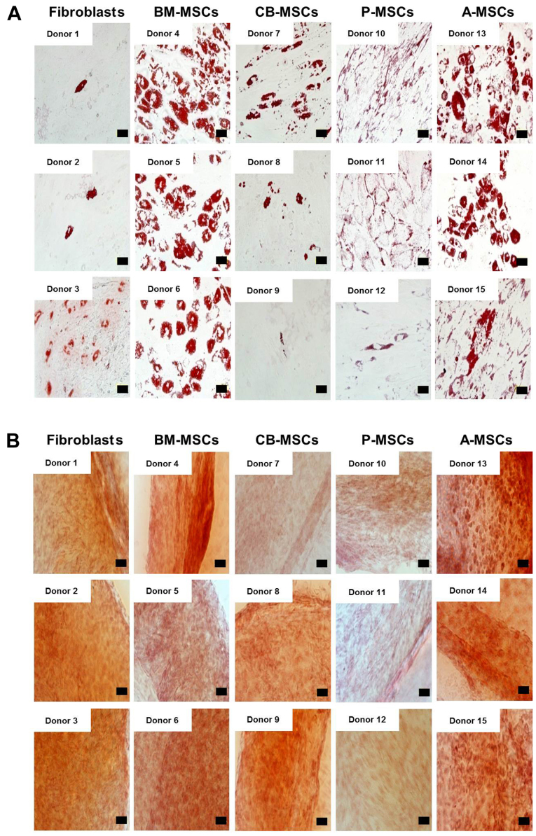
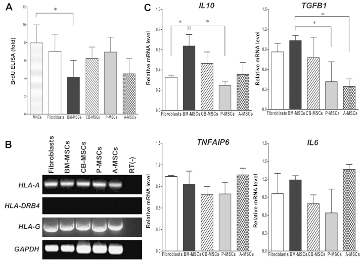
Mesenchymal stem cells (MSCs) are clinically useful due to their capacity for self-renewal, their immunomodulatory properties and tissue regenerative potential. These cells can be isolated from various tissues and exhibit different potential for clinical applications according to their origin, and thus comparative studies on MSCs from different tissues are essential. In this study, we investigated the immunophenotype, proliferative potential, multilineage differentiation and immunomodulatory capacity of MSCs derived from different tissue sources, namely bone marrow, adipose tissue, the placenta and umbilical cord blood. The gene expression profiles of stemness-related genes [octamer-binding transcription factor 4 (OCT4), sex determining region Y-box (SOX)2, MYC, Krüppel-like factor 4 (KLF4), NANOG, LIN28 and REX1] and lineage‑related and differentiation stage-related genes [B4GALNT1 (GM2/GS2 synthase), inhibin, beta A (INHBA), distal-less homeobox 5 (DLX5), runt-related transcription factor 2 (RUNX2), proliferator‑activated receptor gamma (PPARG), CCAAT/enhancer-binding protein alpha (C/EBPA), bone morphogenetic protein 7 (BMP7) and SOX9] were compared using RT-PCR. No significant differences in growth rate, colony-forming efficiency and immunophenotype were observed. Our results demonstrated that MSCs derived from bone marrow and adipose tissue shared not only in vitro tri-lineage differentiation potential, but also gene expression profiles. While there was considerable inter-donor variation in DLX5 expression between MSCs derived from different tissues, its expression appears to be associated with the osteogenic potential of MSCs. Bone marrow-derived MSCs (BM-MSCs) significantly inhibited allogeneic T cell proliferation possibly via the high levels of the immunosuppressive cytokines, IL10 and TGFB1. Although MSCs derived from different tissues and fibroblasts share many characteristics, some of the marker genes, such as B4GALNT1 and DLX5 may be useful for the characterization of MSCs derived from different tissue sources. Collectively, our results suggest that, based on their tri-lineage differentiation potential and immunomodulatory effects, BM-MSCs and adipose tissue-derived MSCs (A-MSCs) represent the optimal stem cell source for tissue engineering and regenerative medicine.


Figures

Figure 1
Figure 1
Growth characteristics and stemness marker expression of human mesenchymal stem cells (MSCs) derived from different tissues. (A) Growth kinetics. Population doubling time (PDT, measured in hours) was determined at each subcultivation. MSCs derived from bone marrow (BM-MSCs), umbilical cord blood (CB-MSCs), the placenta (P-MSCs) and adipose tissue (A-MSCs) that were cultured under identical conditions. (B) Long-term expandability. The finite population doublings, defined as the total number of serial cell passaging before reaching replicative senescence. **p<0.01. (C) Clonogenic capacity was measured by colony forming unit-fibroblast (CFU-F) assay. The results (A–C) were obtained from 3 independent donors and are represented as the means ± SD. (D) Stemness marker expression in MSCs derived from different tissues. RT-PCR analysis for pluripotency markers in MSCs derived from bone marrow, umbilical cord blood, the placenta and adipose tissue compared to induced pluripotent stem (iPS) cell and fibroblasts (F). RT(−) denotes the absence of reverse transcriptase as a control. One representative of 3 independent experiments is shown.
Figure 2
Figure 2
Tri-lineage differentiation of mesenchymal stem cells (MSCs) derived from different tissues. (A) In vitro differentiation assay. MSCs were induced to differentiate toward osteogenic lineage and verified by von Kossa staining after induction (magnification, ×200; scale bar, 100 µm), adipogenic lineage and verified by Oil Red O (magnification, ×400; scale bar, 50 µm), and chondrogenic lineage and verified by Safranin O staining (magnification, ×200; scale bar, 100 µm). One representative of 3 independent experiments is shown. (B) RT-PCR analysis for tri-lineage differentiation-associated markers in MSCs derived from bone marrow (BM-MSCs), umbilical cord blood (CB-MSCs), the placenta (P-MSCs) and adipose tissue (A-MSCs) compared to fibroblasts. The expression of osteogenic (DLX5 and RUNX2), adipogenic (PPARG and C/EBPA) and chondrogenic-associated genes (BMP7 and SOX9) was assayed. The expression of B4GALNT1 was confined to MSCs, and was not noted in fibroblasts. One representative of 3 independent experiments is shown.
Figure 3
Figure 3
(A) Adipogenenic differentiation potential of mesenchymal stem cells (MSCs) derived from different tissue sources. Adipogenic differentiation was carried out for MSCs and fibroblasts isolated from different donors and terminated after 21 days. Fibroblast, bone marrow (BM)-, cord blood (CB)-, placental (P)-, adipose tissue (A)-derived MSCs from different donors were stained by Oil Red O for intracellular lipid vesicles after induction (×400). (Scale bar, 50 µm). (B) Chondrogenic potential of MSCs derived from different tissue sources. Chondrogenic differentiation was induced for 21 days. Fibroblasts, and bone marrow, cord blood, placental, and adipose tissue-derived MSCs from different donors were induced and analyzed by Safranin-O staining (×200 magnification). (Scale bar, 100 µm).
Figure 4
Figure 4
Correlation of DLX5 and osteogenic differentiation capacity of various mesenchymal stem cells (MSCs) from multiple donors. (A) DLX5 transcript of 3 different donors for each MSC derived from different tissues was amplified by RT-PCR. (B) Histologic appearance with von Kossa staining of MSCs of the 3 donors used for RT-PCR in (A). While bone marrow (BM)-derived MSCs and adipose tissue-derived MSCs (A-MSCs) exhibited prominent osteogenic phenotypes, MSCs derived from cord blood and the placenta exhibited inter-donor variation in osteogenic differentiation. (Scale bar, 100 µm).
Figure 5
Figure 5
Immunomodulatory effects of mesenchymal stem cells (MSCs) derived from various sources on activated T cells co-cultured with MSCs. (A) Suppression of human peripheral blood mononuclear cells (MNCs) by MSCs. Proliferation of MNCs (2×10⁵ cells) co-cultured with MSCs (2×10⁴ cells) from different tissues in the presence of 10 µg/ml of phytohaemagglutinin (PHA) for 72 h was evaluated by BrdU ELISA. The data represent the means ± SD of 3 experiments; *p<0.05. (B) Gene expression of HLA-A, HLA-DRB4 and HLA-G for immunomodulation in cells derived from various sources. (C) Relative mRNA expression levels of immunosuppressive RT-PCR of interleukin 10 (IL10), transforming growth factor beta 1 (TGFB1), tumor necrosis factor, alpha-induced protein 6 (TNFAIP6) and IL6 in MSCs from different tissues. Expression levels relative to those of the housekeeping gene, GAPDH are shown. The data represent the means ± SD of 3 experiments; *p<0.05.

References

    1. Jiang Y, Jahagirdar BN, Reinhardt RL, Schwartz RE, Keene CD, Ortiz-Gonzalez XR, Reyes M, Lenvik T, Lund T, Blackstad M, et al. Pluripotency of mesenchymal stem cells derived from adult marrow. Nature. 2002;418:41–49. doi: 10.1038/nature00870. - DOI - PubMed
    1. Petersen BE, Bowen WC, Patrene KD, Mars WM, Sullivan AK, Murase N, Boggs SS, Greenberger JS, Goff JP. Bone marrow as a potential source of hepatic oval cells. Science. 1999;284:1168–1170. doi: 10.1126/science.284.5417.1168. - DOI - PubMed
    1. Prockop DJ, Gregory CA, Spees JL. One strategy for cell and gene therapy: harnessing the power of adult stem cells to repair tissues. Proc Natl Acad Sci USA. 2003;100(Suppl 1):11917–11923. doi: 10.1073/pnas.1834138100. - DOI - PMC - PubMed
    1. Schwartz RE, Reyes M, Koodie L, Jiang Y, Blackstad M, Lund T, Lenvik T, Johnson S, Hu WS, Verfaillie CM. Multipotent adult progenitor cells from bone marrow differentiate into functional hepatocyte-like cells. J Clin Invest. 2002;109:1291–1302. doi: 10.1172/JCI0215182. - DOI - PMC - PubMed
    1. Ryan JM, Barry FP, Murphy JM, Mahon BP. Mesenchymal stem cells avoid allogeneic rejection. J Inflamm (Lond) 2005;2:8. doi: 10.1186/1476-9255-2-8. - DOI - PMC - PubMed

Publication types

MeSH terms