. 2015 Dec 31;9(12):e0004234.
doi: 10.1371/journal.pntd.0004234. eCollection 2015 Dec.

Glycans from Fasciola hepatica Modulate the Host Immune Response and TLR-Induced Maturation of Dendritic Cells

Affiliations

Glycans from Fasciola hepatica Modulate the Host Immune Response and TLR-Induced Maturation of Dendritic Cells

Ernesto Rodríguez et al. PLoS Negl Trop Dis. .

Abstract

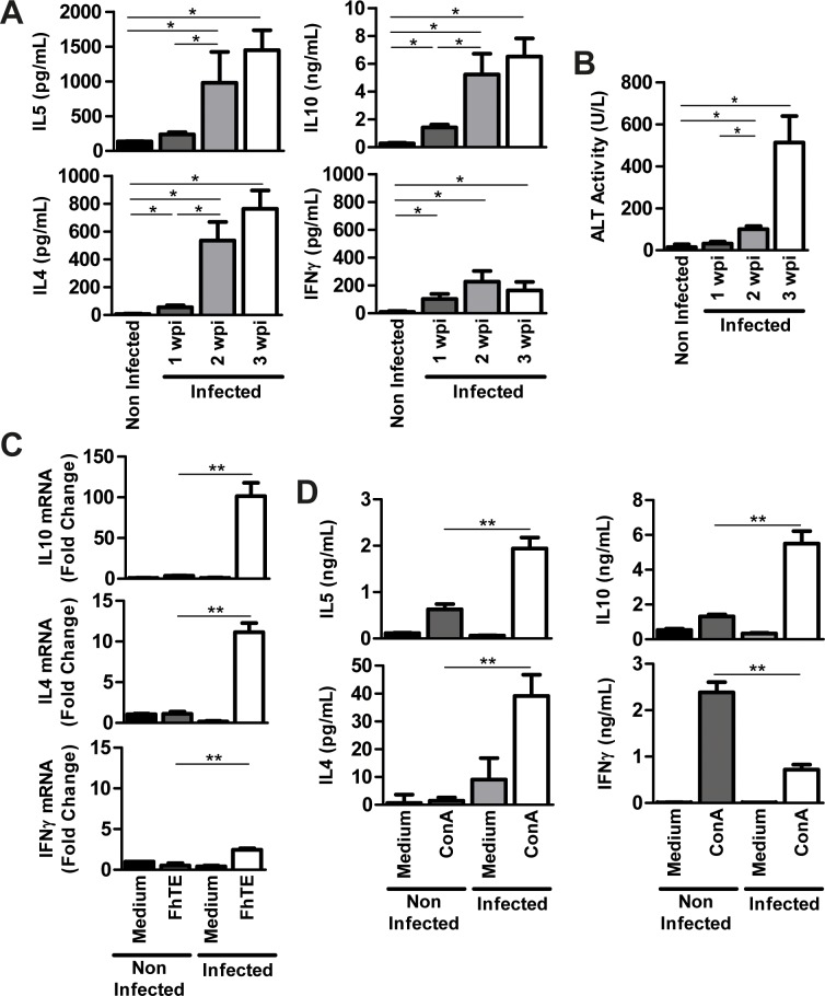
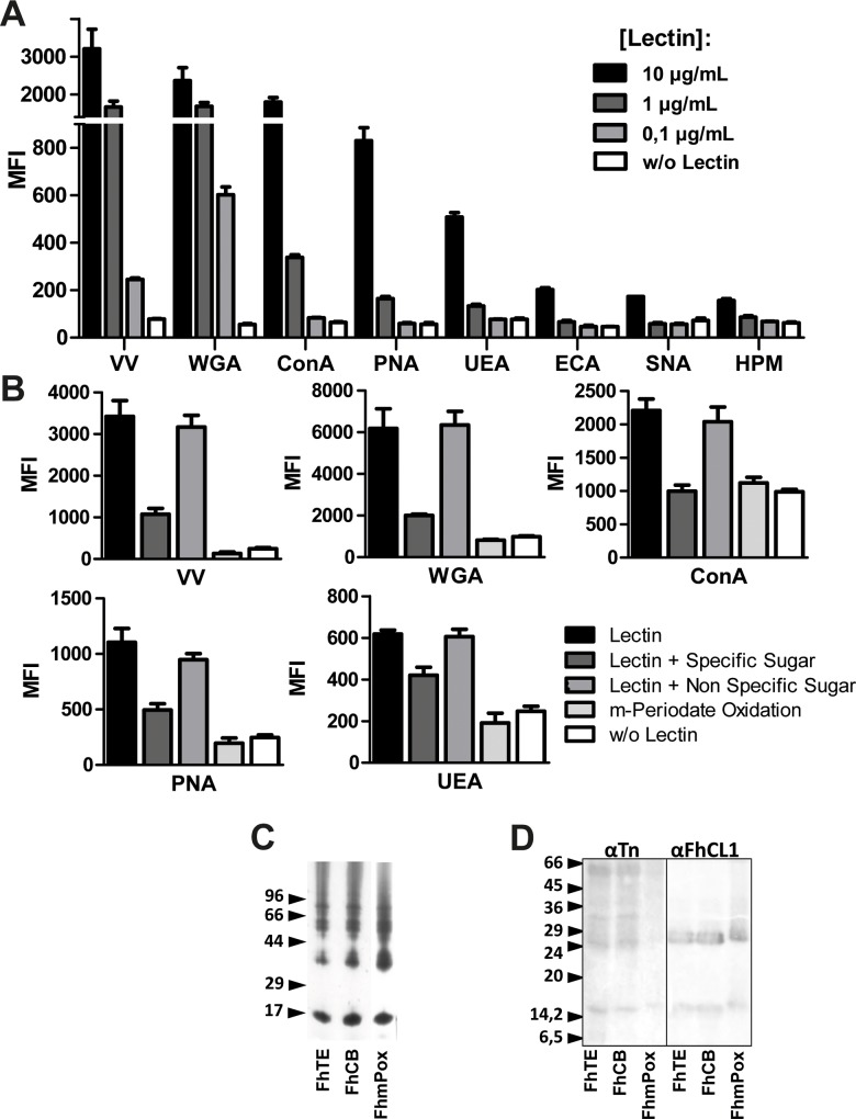
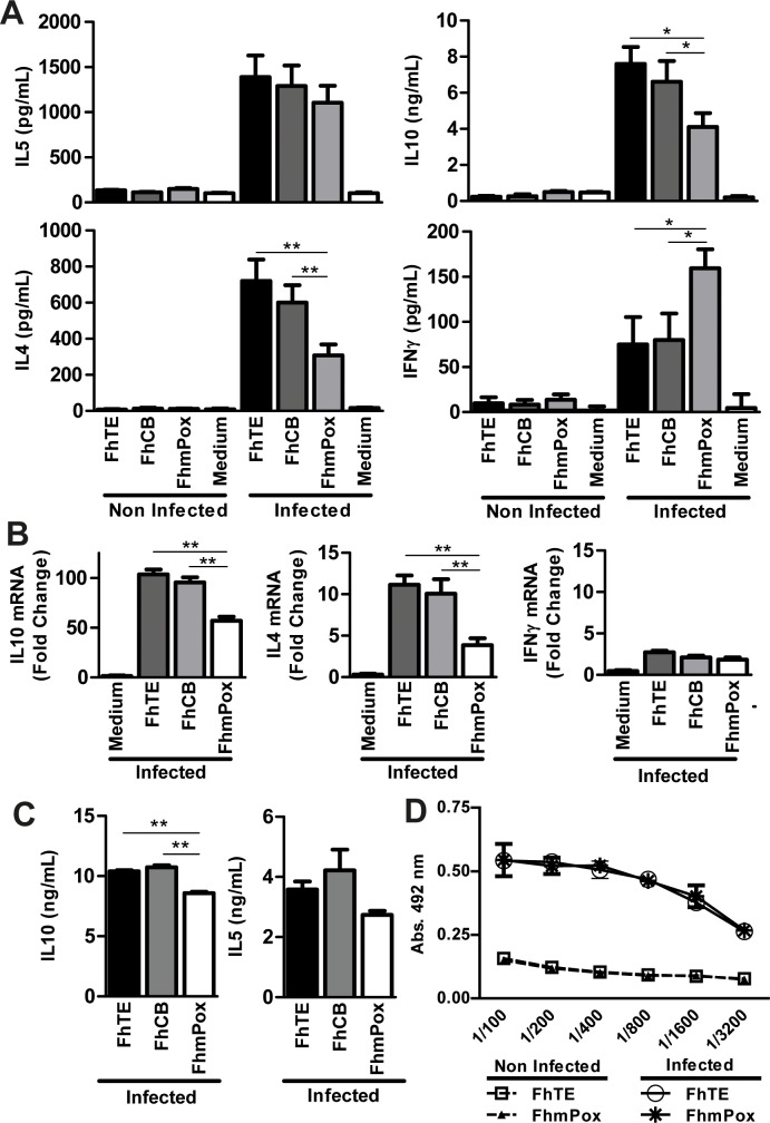
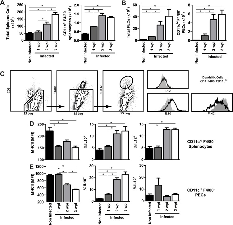
Helminths express various carbohydrate-containing glycoconjugates on their surface, and they release glycan-rich excretion/secretion products that can be very important in their life cycles, infection and pathology. Recent evidence suggests that parasite glycoconjugates could play a role in the evasion of the immune response, leading to a modified Th2-polarized immune response that favors parasite survival in the host. Nevertheless, there is limited information about the nature or function of glycans produced by the trematode Fasciola hepatica, the causative agent of fasciolosis. In this paper, we investigate whether glycosylated molecules from F. hepatica participate in the modulation of host immunity. We also focus on dendritic cells, since they are an important target of immune-modulation by helminths, affecting their activity or function. Our results indicate that glycans from F. hepatica promote the production of IL-4 and IL-10, suppressing IFNγ production. During infection, this parasite is able to induce a semi-mature phenotype of DCs expressing low levels of MHCII and secrete IL-10. Furthermore, we show that parasite glycoconjugates mediate the modulation of LPS-induced maturation of DCs since their oxidation restores the capacity of LPS-treated DCs to secrete high levels of the pro-inflammatory cytokines IL-6 and IL-12/23p40 and low levels of the anti-inflammatory cytokine IL-10. Inhibition assays using carbohydrates suggest that the immune-modulation is mediated, at least in part, by the recognition of a mannose specific-CLR that signals by recruiting the phosphatase Php2. The results presented here contribute to the understanding of the role of parasite glycosylated molecules in the modulation of the host immunity and might be useful in the design of vaccines against fasciolosis.


Conflict of interest statement

The authors have declared that no competing interests exist.

Figures

Fig 1
Fig 1. F. hepatica induces a strong modified-Th2 specific cellular immune response characterized by high levels of IL-4, IL-5 and IL-10.
BALB/c mice (n = 5 per group) were orally infected with 10 metacercariae in PBS (infected mice). PBS alone served as a control (non-infected mice). Mice were bled and sacrificed one, two and three weeks after the infection and spleens were removed. (A) Splenocytes were cultured in the presence of FhTE (75 μg/mL) for 3 days at 37°C. Culture supernatants were collected and analyzed by ELISA for IL-4, IL-5, IL-10 or IFNγ. (B) Alanine transaminase activity was measured in sera from infected and non-infected. (C) Cytokines were also detected by quantitative RT-PCR. Results are shown as the ratio of mRNA amplification for a specific cytokine and the GAPDH. (D) Splenocytes from infected animals sacrificed at 3 wpi and non-infected mice were also stimulated with ConA (5 μg/mL) and cytokines on the supernatants were determined by ELISA. Results are expressed as the mean of three independent experiments (±SD, indicated by error bars). Asterisks indicate statistically significant differences (**p < 0.01; *p < 0.05) with respect to FhTE- or ConA-stimulated splenocytes from non-infected animals.
Fig 2
Fig 2. F. hepatica produces a diverse variety of glycan structures.
(A) Lectin reactivity was evaluated on microplates coated with FhTE (2.5 μg/well) using different concentrations of biotin-conjugated lectins and streptavidin-Dylight-800. Mean fluorescence intensity (MFI) was determined on plates with an infrared imaging system. Lectins used in this study were: VV (GalNAc, Tn antigen), WGA ((GlcNAc)2), ConA (αMan>αGlc), PNA (βGal(1–3)GalNAc) and UEA (Fucα(1–2)Gal), ECA (βGal(1–4)GlcNAc), SNA (αNeuAc(2–6)Gal) and HPM (GalNAc). (B) Carbohydrate specificity was demonstrated by performing inhibition assays with specific carbohydrates (50 mM) by pre-incubating with GalNAc (VV), GlcNAc (WGA), Man (ConA), Gal (PNA) or Fuc (UEA). Alternatively, lectin reactivity was evaluated on periodate-oxidized glycans (FhmPox) or control consisting of FhTE only treated with borydrure (FhCB). (C) Parasite lysates were subjected to SDS-PAGE (15%) and stained with silver nitrate. (D) Alternatively, they were transferred to PVDF membranes and incubated for 2 h at RT with the anti-Tn monoclonal antibody 83D4 or a polyclonal anti-cathepsin L1 serum.
Fig 3
Fig 3. Oxidation of glycans results in a decreased production of IL-4 and IL-10 and an increase of IFNγ by parasite specific splenocytes.
BALB/c mice (n = 5 per group) were infected with 15 metacercariae. After three weeks, animals were bled, sacrificed and spleens and hepatic draining lymph nodes were removed. Splenocytes were cultured in the presence of 75 μg/mL of FhTE, FhCB (oxidation negative control) or FhmPox (oxidized FhTE). Culture supernatants were collected and analyzed by ELISA for IL-4, IL-5, IL-10 or IFNγ (A). Alternatively, IL-4, IL-10 and IFNγ were detected by quantitative RT-PCR. Results are shown as the ratio of mRNA amplification for a specific cytokine and the GAPDH (B). Also, cells from hepatic draining lymph nodes were stimulated with FhTE, FhCB or FhmPox (75 μg/mL) and IL-5 and IL-10 on the supernatants were determined by ELISA (C). Total antibodies in sera from infected and non-infected animals specific for FhTE or FhmPox were evaluated on plates coated with parasite components (10 μg/ml) and with serial dilution of animal sera (D). Results are expressed as the mean of three independent experiments (±SD, indicated by error bars). Asterisks indicate statistically significant differences (**p < 0.01; *p < 0.05) with respect to FhTE-stimulated splenocytes from non-infected animals.
Fig 4
Fig 4. F. hepatica infection promotes the recruitment of MHClow IL-10+ DCs both at the peritoneal cavity and spleen.
Mice (n = 5 per group) were orally infected with 10 metacercariae in PBS (infected mice). PBS alone served as a control (non-infected mice). Mice were sacrificed one, two and three weeks after the infection and spleens and PECs were removed. Splenocyte (A) and PEC (B) suspensions were counted and the presence of CD11chi cells was analyzed by flow cytometry by staining cells with specific antibodies. CD11chi cells were selected after excluding CD3+ followed by exclusion of F4/80+ cells (C). Splenocytes (D) and PECs (E) were also incubated with anti-MCHII, permeabilized, and intracellularly stained with anti-IL-10 and IL-12/23p40 antibodies for 30 min at 4°C. Cells were analyzed on a flow cytometer. Results are expressed as the mean of three independent experiments (±SD, indicated by error bars). Asterisks indicate statistically significant differences (*p < 0.01) with respect to cells from non-infected animals.
Fig 5
Fig 5. BMDCs pulsed with oxidized parasite lysate induce a decreased production of IL-4 and IL-10 by specific CD4+ T cells.
BMDCs were cultured in the presence of 75 μg/mL of FhTE, FhCB (oxidation negative control) or FhmPox (oxidized FhTE) in the presence of LPS (1 μg/ml) overnight at 37°C. Then cell viability was evaluated by incubating with MTT for 4 h and reading absorbance at 570 nm (A). Alternatively, BMDCs were washed twice in complete medium and co-cultured for 3 days at 37°C with total splenocytes (B) or purified CD4+ T cells (C) from infected animals sacrificed after 3 wpi. Culture supernatants were collected and analyzed by ELISA for IL-4, IL-5, IL-10 or IFNγ Results are expressed as the mean of three independent experiments (±SD, indicated by error bars). Asterisks indicate statistically significant differences (**p < 0.01; *p < 0.05) with respect to FhTE/LPS-stimulated DCs.
Fig 6
Fig 6. Glycoconjugates from F. hepatica interact with and are uptaken by BMDCs in a process mediated by Man-specific CLRs.
BMDCs were incubated with Alexa 647-labeled FhTE for 1 h at 37°C in complete medium (to assess uptake), or at 4°C in complete medium (to assess binding) in presence or absence of EDTA (5 mM) and analyzed by flow cytometry on CD11c+ cells (A). Atto-labeled-FhTE, FhCB (control) or FhmPox (oxidized FhTE) were also incubated with BMDC and analyzed in the same conditions as in A (B). Inhibition assays with carbohydrates were carried out on BMDCs pre-incubated with 50 mM of different carbohydrates or laminarin for 30 min (C). Asterisks indicate statistically significant differences (p < 0.01) with respect to BMDC incubated with FhTE.
Fig 7
Fig 7. Oxidation of parasite components partially inhibits modulation of LPS-induced maturation of DCs by FhTE.
BMDCs were cultured in the presence of 75 μg/mL of FhTE, FhCB (oxidation negative control) or FhmPox (oxidized FhTE) in presence or absence of LPS (1 μg/ml) overnight at 37°C. Then, culture supernatants were collected and analyzed by ELISA for IL-6, IL-10 or IL-12/23p40 (A). BMDCs were also pre-incubated for 45 min. with mannose (Man), N-Acetyl-Galactosamine (GalNAc) or arabinose (Ara) at 10 mM and then stimulated as in A. Culture supernatants were analyzed by ELISA for detection of IL-6, IL-10 or IL-12/23p40 (B) and MIP-1α and MIP-2 (C). Alternatively, BMDCs were pre-incubated for 45 min. with 10 μM of specific signaling inhibitors (PHPS1; GW5074; and ER27319) and then stimulated with FhTE (75 μg/mL) in presence of LPS (1 μg/ml) (D). Results are expressed as the mean of three independent experiments (±SD, indicated by error bars). Asterisks indicate statistically significant differences (*p < 0.01) with respect to LPS-stimulated BMDCs.

Similar articles

Cited by

References

    1. Rojo-Vázquez F, Meana A, Valcárcel F, Martínez-Valladares M. Update on trematode infections in sheep. Vet Parasitol. 2012;189:15–38. 10.1016/j.vetpar.2012.03.029 - DOI - PubMed
    1. Toet H, Piedrafita DM, Spithill TW. Liver fluke vaccines in ruminants: strategies, progress and future opportunities. Int J Parasitol. 2014. October 15;44(12):915–27. 10.1016/j.ijpara.2014.07.011 - DOI - PubMed
    1. Flynn R, Mulcahy G. The roles of IL-10 and TGF-beta in controlling IL-4 and IFN-gamma production during experimental Fasciola hepatica infection. Int J Parasitol. 2008;38:1673–80. 10.1016/j.ijpara.2008.05.008 - DOI - PubMed
    1. Walsh K, Brady M, Finlay C, Boon L, Mills K. Infection with a helminth parasite attenuates autoimmunity through TGF-beta-mediated suppression of Th17 and Th1 responses. J Immunol. 2009;183:1577–86. 10.4049/jimmunol.0803803 - DOI - PubMed
    1. Dowling D, Hamilton C, Donnelly S, La Course J, Brophy P, Dalton J, et al. Major secretory antigens of the helminth Fasciola hepatica activate a suppressive dendritic cell phenotype that attenuates Th17 cells but fails to activate Th2 immune responses. Infect Immun. 2010;78:793–801. 10.1128/IAI.00573-09 - DOI - PMC - PubMed

Publication types

LinkOut - more resources