. 2015 Dec 31;10(12):e0141925.
doi: 10.1371/journal.pone.0141925. eCollection 2015.

Robo 4 Counteracts Angiogenesis in Herpetic Stromal Keratitis

Affiliations

Robo 4 Counteracts Angiogenesis in Herpetic Stromal Keratitis

Fernanda Gimenez et al. PLoS One. .

Abstract

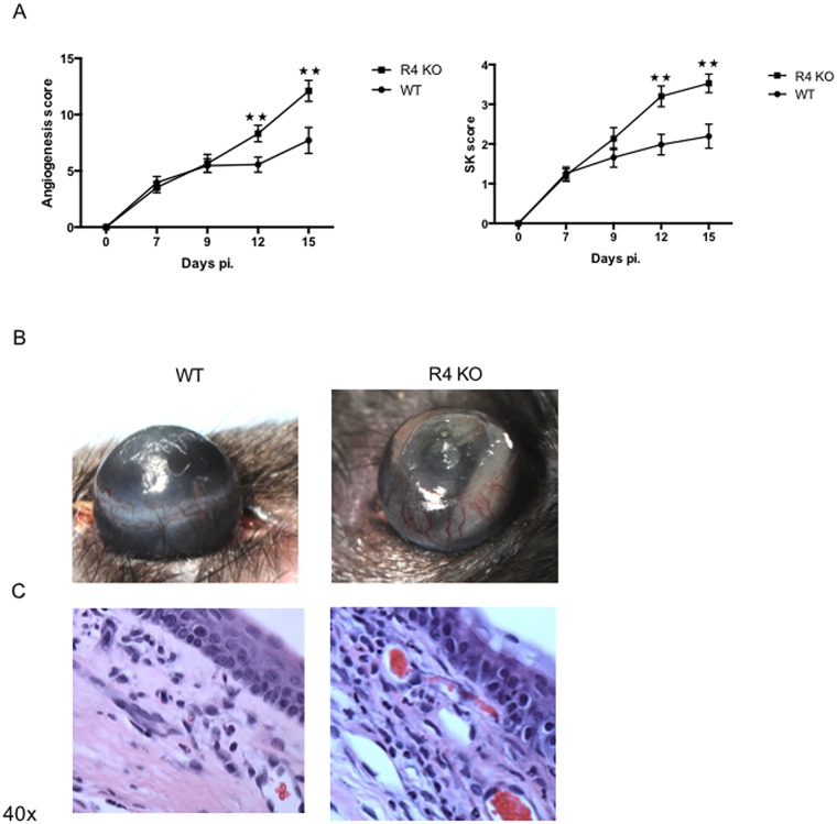
The cornea is a complex tissue that must preserve its transparency to maintain optimal vision. However, in some circumstances, damage to the eye can result in neovascularization that impairs vision. This outcome can occur when herpes simplex virus type 1 (HSV-1) causes the immunoinflammatory lesion stromal keratitis (SK). Potentially useful measures to control the severity of SK are to target angiogenesis which with herpetic SK invariably involves VEGF. One such way to control angiogenesis involves the endothelial receptor Robo4 (R4), which upon interaction with another protein activates an antiangiogenic pathway that counteracts VEGF downstream signaling. In this study we show that mice unable to produce R4 because of gene knockout developed significantly higher angiogenesis after HSV-1 ocular infection than did infected wild type (WT) controls. Moreover, providing additional soluble R4 (sR4) protein by subconjunctival administration to R4 KO HSV-1 infected mice substantially rescued the WT phenotype. Finally, administration of sR4 to WT HSV-1 infected mice diminished the extent of corneal angiogenesis compared to WT control animals. Our results indicate that sR4 could represent a useful therapeutic tool to counteract corneal angiogenesis and help control the severity of SK.


Conflict of interest statement

Competing Interests: The authors have declared that no competing interests exist.

Figures

Fig 1
Fig 1. Robo4 deficient mice are more susceptible to HSV-1 infection.
R4 KO mice were infected with HSV-1 RE. (A) SK lesion and angiogenesis severity were significantly increased in R4 KO mice compared to WT mice on day 15 pi. (B) Representative eye photos show increased SK lesion and angiogenesis severity in R4 KO compared to WT mice (C) Eyes were processed for cryo-sections on day 15 pi. Hematoxilyn and eosin staining was carried out on 6 μm sections, and pictures were taken at 40x magnification. The sections show decreased cellular infiltration in R4 KO mice compared to WT mice. Data are representative of three independent experiments and show mean values ± SEM (n = 15 mice/group). ***p ≤ 0.001, **p ≤ 0.01, *p ≤ 0.05. Statistical levels of significance were analyzed by t test.
Fig 2
Fig 2. Robo4 deficient mice present more inflammation and vascularization.
Robo4 knockout (R4 KO) and WT mice were infected with HSV-1 RE and at day 15 pi. corneas were collected and pooled for analysis by flow cytometry or Q-RT-PCR. The frequency and total cell number per cornea for (A) endothelial cells (CD31+) gated on total CD45- cells infiltrate, (B) CD4+ T cells (CD4+) (gated on total CD45+ cells infiltrate) and (C) neutrophils (Ly6G+ CD11b+ gated on total CD45+ cells infiltrate) show significant increase in R4 KO mice. Data are a combination of 3 independent experiments and show mean values ± SEM (n = 7 and each sample is representative of 2 corneas). ***p ≤ 0.001, **p ≤ 0.01, *p ≤ 0.05. Statistical levels of significance were analyzed by t test. (E) Relative fold change in mRNA expression of IL-1β, IL-6 and CXCL-1 was examined and compared between WT and Robo4 KO mice on day 15 pi. by Q-RT-PCR. Data represent means ± SEM from two different independent experiments (n = 3 and each sample is representative of 5 corneas). ***p ≤ 0.001, **p ≤ 0.01, *p ≤ 0.05. Statistical levels of significance were analyzed by t test.
Fig 3
Fig 3. Administration of soluble Robo4 (sR4) shifts the Robo4 knockout to the WT phenotype.
WT and Robo4 knockout mice (R4 KO) were infected with HSV-1 RE. (A) R4 KO mice received either sR4 (R4 KO treated w/ sR4) or vehicle (R4 KO Control) from 2 to 14 days pi. WT mice were included and received vehicle (WT Control) under the same regimen previously stated. (B) SK lesions and angiogenesis severity was decreased in R4 KO mice treated with sR4. (C) Representative eye photos show that R4 KO mice treated with sR4 do not develop the severe phenotype that R4 KO control animals do (D) Hematoxylin and eosin staining was carried out on 6-μm sections, and pictures were taken 40 x magnification. Representative eye sections show decreased cellular infiltration in R4 KO treated w/ sR4 and WT control compared to R4 KO control mice. Data are representative of two independent experiments and show mean values ± SEM (n = 12 mice/group). ***p ≤ 0.001,**p ≤ 0.01,*p ≤ 0.05. Statistical levels of significance were analyzed by one-way ANOVA test with Tuckey’s post hoc test settings.
Fig 4
Fig 4. Preventive administration of soluble Robo4 (sR4) reduces lesion severity in HSV-1 infected mice.
WT mice were infected with HSV-1 RE and treated with sR4 or vehicle (PBS) (A) The sR4 treatment was given to HSV-1 infected mice as shown (B) Dose dependent inhibition of angiogenesis scores after sR4 treatment. Data represents means ± SEM from three different experiments (n = 10) (C) sR4 treatment regimen resulted in SK and angiogenesis scores reduction. Data are representative of three independent experiments and show mean values ± SEM (n = 15 mice/group). ***p ≤ 0.001, **p ≤ 0.01, *p ≤ 0.05. Statistical levels of significance were analyzed by t test. (D) Representative eye photos show decreased SK lesion and angiogenesis severity in sR4 treated mice compared to control mice (E) Eyes were processed for cryo-sections on day 15 pi. Hematoxilyn and eosin staining was carried out on 6 μm sections, and pictures were taken at 40x magnification. The sections show decreased cellular infiltration in mice treated with sR4 compared to control mice.
Fig 5
Fig 5. Provision of sR4 reduces corneal inflammation and angiogenesis.
WT mice were infected with HSV-1 RE and treated with sR4 of vehicle (PBS) from day 2 pi. to 14 pi. At day 15 pi. corneas were collected and pooled for analysis by flow cytometry or Q-RT-PCR. The frequency and total cell number per cornea for (A) endothelial cells (CD31+) gated on total CD45- cells infiltrate, (B) CD4+ T cells (CD4+) (gated on total CD45+ cells infiltrate) and (C) neutrophils (Ly6G+ CD11b+ gated on total CD45+ cells infiltrate) show significant decrease in sR4 treated mice compared to WT mice. Data are a combination of 3 independent experiments and show mean values ± SEM (n = 7 and each sample is representative of 2 corneas). ***p ≤ 0.001, **p ≤ 0.01, *p ≤ 0.05. Statistical levels of significance were analyzed by t test. (D) Relative fold change in mRNA expression of IL-1β, IL-6 and CXCL-1 was examined and compared between sR4 treated and control mice on day 15 pi. by Q-RT-PCR. Data represent means ± SEM from three different independent experiments (n = 2 and each sample is representative of 6 corneas). ***p ≤ 0.001, **p ≤ 0.01, *p ≤ 0.05. Statistical levels of significance were analyzed by t test.
Fig 6
Fig 6. Increased lesion severity in Robo4 KO mice is due to higher VEGF levels and signaling.
WT and R4 KO mice were infected with HSV-1 RE in one eye and corneas were collected and pooled for analysis by WB and by Q-RT-PCR. (A) Relative fold change in mRNA expression of VEGF was examined and compared between WT and R4 KO animals. Data represent means ± SEM (n = 2 and each sample is representative of 6 corneas). ***p ≤ 0.001, **p ≤ 0.01, *p ≤ 0.05. Statistical levels of significance were analyzed by t test. (B) WB analysis of corneal lysates. R4 KO animals have increased VEGF signaling showed by increased SRC phosphorylation at day 15 pi. (n = 3 and each sample is representative of 5 corneas).

References

    1. Niederkorn JY (2006) See no evil, hear no evil, do no evil: the lessons of immune privilege. Nat Immunol 7: 354–359. - PubMed
    1. Streilein JW (2003) Ocular immune privilege: the eye takes a dim but practical view of immunity and inflammation. J Leukoc Biol 74: 179–185. - PubMed
    1. Liesegang TJ (2001) Herpes simplex virus epidemiology and ocular importance. Cornea 20: 1–13. - PubMed
    1. Giménez F, Suryawanshi A, Rouse BT (2013) Pathogenesis of herpes stromal keratitis—a focus on corneal neovascularization. Prog Retin Eye Res 33: 1–9. 10.1016/j.preteyeres.2012.07.002 - DOI - PMC - PubMed
    1. Suryawanshi A, Mulik S, Sharma S, Reddy PB, Sehrawat S, et al. (2011) Ocular neovascularization caused by herpes simplex virus type 1 infection results from breakdown of binding between vascular endothelial growth factor A and its soluble receptor. J Immunol 186: 3653–3665. 10.4049/jimmunol.1003239 - DOI - PMC - PubMed

Publication types

MeSH terms

Substances