. 2015 Dec 31;6(12):e2026.
doi: 10.1038/cddis.2015.375.

Myocyte-derived Tnfsf14 is a survival factor necessary for myoblast differentiation and skeletal muscle regeneration

Affiliations

Myocyte-derived Tnfsf14 is a survival factor necessary for myoblast differentiation and skeletal muscle regeneration

R J Waldemer-Streyer et al. Cell Death Dis. .

Abstract

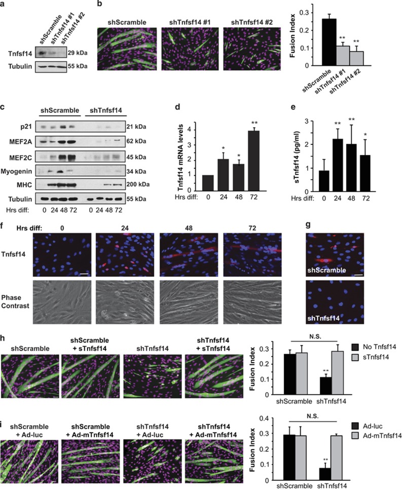
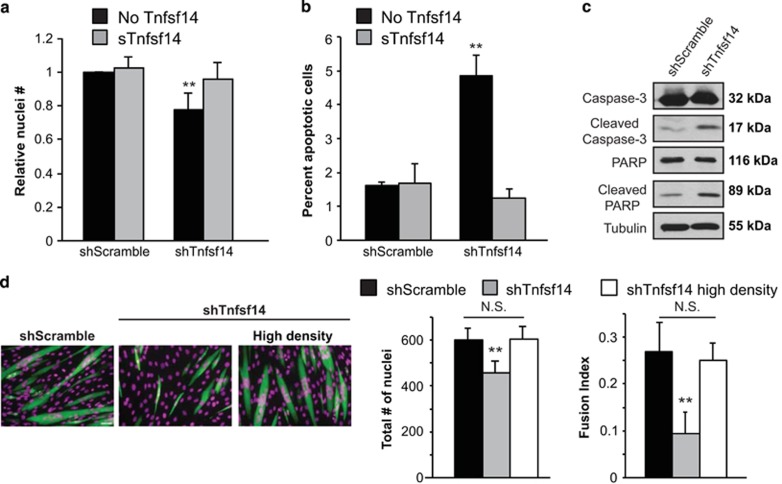
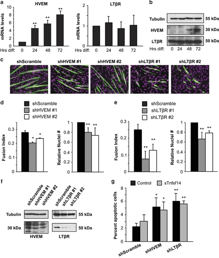
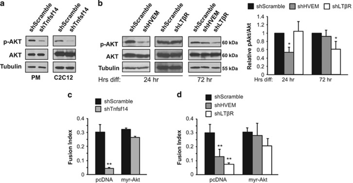
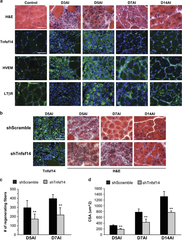
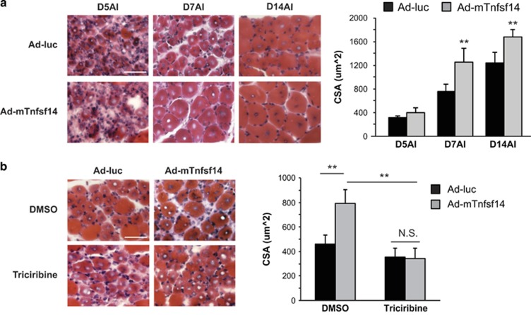
Adult skeletal muscle tissue has a uniquely robust capacity for regeneration, which gradually declines with aging or is compromised in muscle diseases. The cellular mechanisms regulating adult myogenesis remain incompletely understood. Here we identify the cytokine tumor necrosis factor superfamily member 14 (Tnfsf14) as a positive regulator of myoblast differentiation in culture and muscle regeneration in vivo. We find that Tnfsf14, as well as its cognate receptors herpes virus entry mediator (HVEM) and lymphotoxin β receptor (LTβR), are expressed in both differentiating myocytes and regenerating myofibers. Depletion of Tnfsf14 or either receptor inhibits myoblast differentiation and promotes apoptosis. Our results also suggest that Tnfsf14 regulates myogenesis by supporting cell survival and maintaining a sufficient pool of cells for fusion. In addition, we show that Akt mediates the survival and myogenic function of Tnfsf14. Importantly, local knockdown of Tnfsf14 is found to impair injury-induced muscle regeneration in a mouse model, affirming an important physiological role for Tnfsf14 in myogenesis in vivo. Furthermore, we demonstrate that localized overexpression of Tnfsf14 potently enhances muscle regeneration, and that this regenerative capacity of Tnfsf14 is dependent on Akt signaling. Taken together, our findings reveal a novel regulator of skeletal myogenesis and implicate Tnfsf14 in future therapeutic development.


Figures

Figure 1
Figure 1
Tnfsf14 is a positive regulator of myoblast differentiation. (a) C2C12 cells were infected with lentiviruses expressing shTnfsf14 or shScramble (negative control), selected for 2 days, followed by cell lysis and western analysis (n=4). (b) Cells treated as in (a) were differentiated for 72 h, followed by staining for MHC (green) and DAPI (pseudo-colored magenta), and quantification of fusion index (n=5). (c) Cells treated as in (a) were differentiated, and at indicated time points ('Hrs diff') were lysed and subjected to western analysis (n=4). (d) RNA was isolated from differentiating C2C12 cells at indicated time points and subjected to qRT-PCR analysis for Tnfsf14 mRNA levels (n=6). (e) Cell media over the course of differentiation were subjected to ELISA assay to determine sTnfsf14 levels (n=8). (f) Differentiating C2C12 cells at various time points ('Hrs diff') were stained without permeabilization for Tnfsf14 (red) and DAPI (blue) (n=4). (g) Cells treated as in a were differentiated for 24 h, followed by staining for Tnfsf14 (red) and DAPI (blue) (n=3). (h) C2C12 cells were treated as in (a), and then differentiated in the presence or absence of 25 ng/ml recombinant sTnfsf14 for 3 days, followed by staining for MHC and DAPI, and quantification of fusion index (n=3). (i) C2C12 cells were treated as in (a), and then infected with adenoviruses expressing mTnfsf14 or luciferease (luc; negative control), followed by differentiation for 3 days and then staining for MHC and DAPI. The fusion index was quantified (n=3). All error bars represent S.D. of independent replicates. One-sample two-tailed t-test was performed for data in (d), and paired two-tailed t-tests for all other data. *P<0.05; **P<0.01. Scale bars: 50 μm
Figure 2
Figure 2
Tnfsf14 maintains sufficient cell numbers for differentiation. (a) C2C12 cells were infected with lentiviruses expressing shTnfsf14 or shScramble, selected for 2 days, followed by differentiation for 72 h in the presence or absence of 25 ng/ml recombinant sTnfsf14 and subsequent staining with DAPI. Stained nuclei were counted (n=3). (b) Cells were treated as in (a) but differentiated for 24 h, followed by TUNEL assay to detect apoptotic cells (n=4). (c) Primary myoblasts were infected with shRNA lentiviruses and differentiated for 24 h, followed by cell lysis and western analysis (n=3). (d) C2C12 cells were infected with shRNA lentiviruses and then plated at different densities in order to compensate for the lower cell number seen in Tnfsf14 knockdown cells. Cells were differentiated for 72 h and subsequently stained for MHC. Nuclei number and fusion index were quantified (n=4). Scale bar: 50 μm. One-sample two-tailed t-test was performed for data in (a), and paired two-tailed t-tests for all other data. **P<0.01. All error bars represent S.D. of independent replicates
Figure 3
Figure 3
Both HVEM and LTβR are required for myoblast differentiation and survival. (a) HVEM or LTβR mRNA transcripts were measured over the course of C2C12 differentiation by qRT-PCR (n=4 for HVEM and n=3 for LTβR). (b) HVEM and LTβR protein levels during C2C12 cell differentiation were analyzed by western blotting (n=4). (c) C2C12 cells were infected with shRNA lentiviruses as indicated, selected for 2 days, followed by differentiation for 72 h and subsequent staining for MHC and with DAPI. Scale bar: 50 μm. (d and e) Fusion index and total nuclei number were quantified for experiments in (c) (n=5). (f) Lentivirus-mediated knockdown of HVEM and LTβR in C2C12 cells were confirmed by western analysis (n=3 for HVEM and n=4 for LTβR). (g) C2C12 cells treated as in (c) but differentiated for 24 h were subjected to TUNEL assays, and the percentage of apoptotic cells was measured (n=4). One-sample two-tailed t-test was performed for data in (a) and relative nuclei number in (d) and (e), paired two-tailed t-tests for all other data. *P<0.05; **P<0.01. All error bars represent S.D. of independent replicates
Figure 4
Figure 4
Tnfsf14 regulates myoblast differentiation through Akt. (a) Primary mouse myoblasts (n=4) and C2C12 cells (n=5) were infected with shRNA lentiviruses and differentiated for 24 h, followed by cell lysis and western analysis. (b) C2C12 cells were infected with shRNA lentiviruses as indicated and differentiated for 24 or 72 h, followed by cell lysis and western analysis (n=5). (c and d) C2C12 cells were infected with shRNA lentiviruses as indicated, and then transfected with a constitutively active Akt or a control (pcDNA) construct, followed by differentiation for 72 h and subsequent staining for MHC and with DAPI. Fusion index and total nuclei number were quantified (n=4). One-sample two-tailed t-test was performed for data in (b) and paired two-tailed t-tests for data in (c) and (d). *P<0.05; **P<0.01. All error bars represent S.D. of independent replicates
Figure 5
Figure 5
Tnfsf14 is required for robust muscle regeneration in vivo. (a) TA muscles were injured by BaCl2 injection, and isolated on days 3, 5, 7 and 14 AI. Upon cryosection, H&E staining or immunofluorescence staining for Tnfsf14, HVEM and LTβR (green) together with DAPI (blue) were performed (n=3). (b) TA muscles were co-injected with BaCl2 and shRNA viruses, and then processed as described in (a). (c) Quantification of the number of regenerating myofibers on muscle sections from (b). (d) Quantification of the regenerating myofiber cross-sectional area (CSA) on muscle sections from (b). For each time point in (bd), five or six mice were analyzed. All error bars represent S.D. of independent replicates. Paired two-tailed t-test was performed to compare shScramble and shTnfsf14 at each time point. **P<0.01. Scale bars: 50 μm
Figure 6
Figure 6
Local overexpression of Tnfsf14 enhances skeletal muscle regeneration in an Akt-dependent manner. (a) TA muscles were co-injected with BaCl2 and adenoviruses expressing mTnfsf14 (Ad-mTnfsf14) or luciferase (Ad-luc), and isolated on days 5, 7 and 14 AI. Upon cryosection, H&E staining was performed, and regenerating myofiber CSA was quantified. For each time point, five to seven mice were analyzed. (b) The procedure described in a was repeated, and the animals received daily intraperitoneal injection of 100 μl triciribine (0.26 μg/μl) or 20% DMSO as a control, starting on the day of BaCl2 injection. For each condition, six mice were analyzed. All error bars represent S.D. of independent replicates. Paired two-tailed t-test was performed. **P<0.01. Scale bars: 50 μm

References

    1. 1Relaix F, Marcelle C. Muscle stem cells. Curr Opin Cell Biol 2009; 21: 748. - PubMed
    1. 2Tidball JG. Mechanisms of muscle injury, repair, and regeneration. Compr Physiol 2011; 1: 2029. - PubMed
    1. 3Naya FS, Olson E. MEF2: a transcriptional target for signaling pathways controlling skeletal muscle growth and differentiation. Curr Opin Cell Biol 1999; 11: 683. - PubMed
    1. 4Parker MH, Seale P, Rudnicki MA. Looking back to the embryo: defining transcriptional networks in adult myogenesis. Nat Rev Genet 2003; 4: 497. - PubMed
    1. 5Wagers AJ, Conboy IM. Cellular and molecular signatures of muscle regeneration: current concepts and controversies in adult myogenesis. Cell 2005; 122: 659. - PubMed

Publication types

Substances