. 2015 Dec;56(13):8258-67.
doi: 10.1167/iovs.15-17073.

Generating iPSC-Derived Choroidal Endothelial Cells to Study Age-Related Macular Degeneration

Affiliations

Generating iPSC-Derived Choroidal Endothelial Cells to Study Age-Related Macular Degeneration

Allison E Songstad et al. Invest Ophthalmol Vis Sci. 2015 Dec.

Abstract

Purpose: Age-related macular degeneration (AMD), the most common cause of incurable blindness in the western world, is characterized by the dysfunction and eventual death of choroidal endothelial (CECs), RPE, and photoreceptor cells. Stem cell-based treatment strategies designed to replace photoreceptor and RPE cells currently are a major scientific focus. However, the success of these approaches likely also will require replacement of the underlying, supportive choroidal vasculature. The purpose of this study was to generate stem cell-derived CECs to develop efficient differentiation and transplantation protocols.

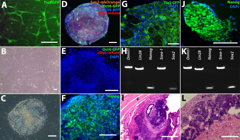
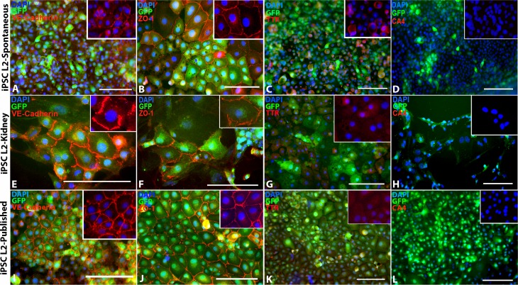
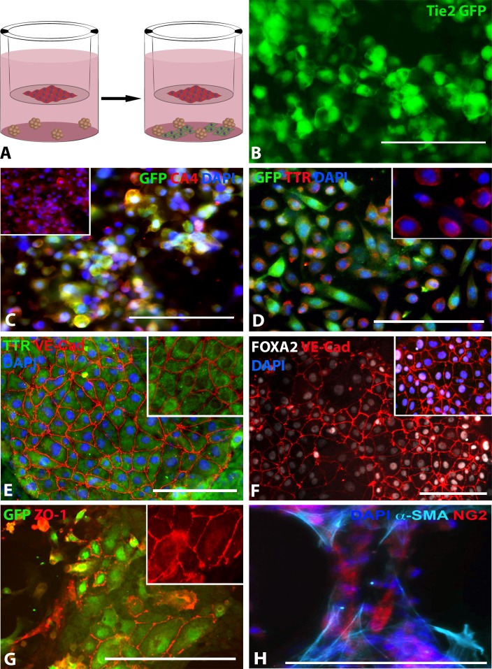
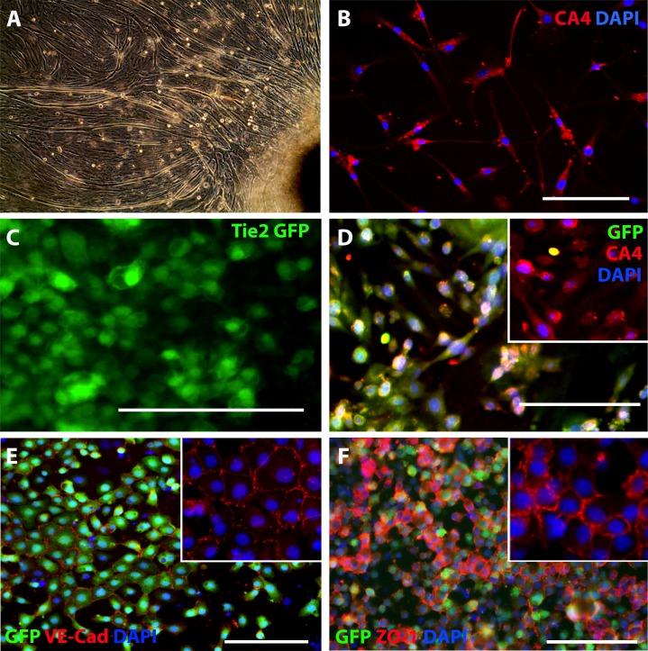
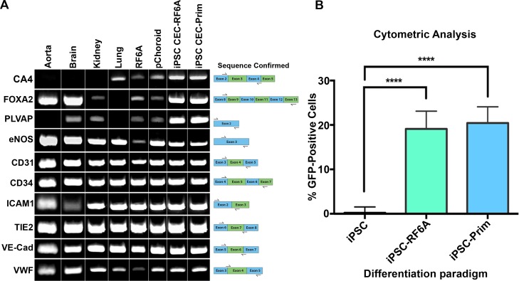
Methods: Dermal fibroblasts from the Tie2-GFP mouse were isolated and reprogrammed into two independent induced pluripotent stem cell (iPSC) lines via viral transduction of the transcription factors Oct4, Sox2, Klf4, and c-Myc. Tie2-GFP iPSCs were differentiated into CECs using a coculture method with either the RF6A CEC line or primary mouse CECs. Induced pluripotent stem cell-derived CECs were characterized via RT-PCR and immunocytochemistry for EC- and CEC-specific markers.

Results: Induced pluripotent stem cells generated from mice expressing green fluorescent protein (GFP) under control of the endothelial Tie2 promoter display classic pluripotency markers and stem cell morphology. Induced pluripotent stem cell-derived CECs express carbonic anhydrase IV, eNOS, FOXA2, PLVAP, CD31, CD34, ICAM-1, Tie2, TTR, VE-cadherin, and vWF.

Conclusions: Induced pluripotent stem cell-derived CECs will be a valuable tool for modeling of choriocapillaris-specific insults in AMD and for use in future choroidal endothelial cell replacement approaches.


Figures

Figure 1
Figure 1
Reprogramming Tie2 GFP fibroblasts into iPSCs. (A) Green fluorescence protein expression in Tie2-GFP heart and lung primary ECs. (B) Tail-tip fibroblasts were isolated from Tie2-GFP mice and reprogrammed into (C) iPSCs via transduction with either (D) fluorescently tagged lentiviral vectors (iPSC-L1) or a (J) polycistronic lentiviral vector (iPSC-L2). (E) The FACS iPSC-L1 cells did not express the exogenous fluorescent tags eGFP-Oct4 and mKate-c-Myc (4′,6-diamidino-2-phenylendole [DAPI] = blue), but did express (F) Nanog, indicating the iPSC-L1 cells were fully reprogrammed. (G) iPSC-L1 EBs expressed GFP (green), indicating the presence of ECs (DAPI = blue). (H, K) Reverse transcription-PCR analysis of pluripotency markers in both iPSC lines (iPSC-L1 and iPSC-L2, respectively). (J) Nanog expression (green) in iPSC-L2 iPSCs (DAPI = blue). (I, L) Teratoma formation assays for iPSC-L1 and iPSC-L2, respectively. ∧ = endoderm, * = ectoderm, + = mesoderm). Scale bars: 100 μm.
Figure 2
Figure 2
Spontaneous iPSC-EC differentiation, kidney coculture iPSC-EC differentiation, and previously published iPSC- EC differentiation protocol. (A–D) Spontaneously differentiated iPSC-derived ECs express EC markers (A) VE-Cadherin and (B) ZO-1, a small amount of (C) TTR, but no (D) CA4. (EH) iPSC-derived ECs differentiated in coculture with primary mouse kidney ECs also express (E) VE-Cadherin, (F) ZO-1, and (G) TTR, but not (H) CA4. (IL) Likewise, iPSC-derived ECs differentiated using a previously published iPSC-EC differentiation protocol from Rufaihah et al. also express (I) VE-Cadherin, (J) ZO-1, and (K) TTR, but not (L) CA4. DAPI = blue. Scale bars: 100 μm.
Figure 3
Figure 3
Differentiating choroidal-like ECs via co-culture with RF6A CECs. (A) Schematic illustrating differentiation paradigm: red cells = RF6A CECs, green cells = differentiated iPSC-ECs. (B) GFP expression in live iPSC-L2-RF6A ECs. (C) Carbonic anhydrase IV (CA4; red), GFP (green), and DAPI (blue) expression in iPSC-L2-RF/6A ECs; inset of only CA4 and DAPI expression. (D) GFP (green), TTR (red), and DAPI (blue) expression in iPSC-L2-RF/6A ECs, inset of only TTR and DAPI expression. (E) TTR (green), VE-Cadherin (red), and DAPI (blue) expression in iPSC-L2-RF/6A ECs; inset of only TTR and VE-Cadherin expression; pseudocoloring performed using Fiji. (F) Green fluorescent protein (green) and ZO-1 (red) expression in iPSC-L2-RF/6A ECs; inset of only ZO-1 expression. (G) FOXA2 (gray), VE-Cadherin (red), and DAPI (blue) expression in iPSC-L2-RF/6A ECs; pseudocoloring performed using Fiji. (H) α-SMA (cyan), NG-2 (red), and DAPI expression in iPSC-L2-RF/6A pericytes; pseudocoloring performed using Fiji. Scale bars: 100 μm.
Figure 4
Figure 4
Differentiating choroidal-like ECs via coculture with primary CECs. (A, F) Isolated primary CECs from mouse choroids (CA4 = red, DAPI = blue). (GJ) iPSC-L2 differentiated in coculture with primary CECs. (B, G) Green fluorescent protein expression in live iPSC-L2-Prim ECs. (C, H) Carbonic anhydrase IV (CA4; red), GFP (green), and DAPI (blue) expression in iPSC-L2-Prim ECs; insets of CA4 and DAPI expression. (D, I) Green fluorescent protein (green), VE-Cadherin (red), and DAPI (blue) expression in iPSC-L2-Prim ECs. (E, J) Green fluorescent protein (green), ZO-1 (red), and DAPI (blue) expression in iPSC-L2-Prim ECs. Scale bars: 100 μm.
Figure 5
Figure 5
Comparing EC-specific marker expression in iPSC-ECs to other ECs and quantifying percentage of GFP-positive cells in iPSC-ECs. (A) Reverse transcription-PCR analysis of various EC markers in different cell types. (B) Tali cytometer analysis of iPSC-L2-RF6A and iPSC-L2-Prim lines compared to iPSC L2. **P ≤ 0.01; ***P ≤ 0.001; ****P ≤ 0.0001.

References

    1. Buch H,, Vinding T,, La Cour M,, Nielsen NV. The prevalence and causes of bilateral and unilateral blindness in an elderly urban Danish population. The Copenhagen City Eye Study. Acta Ophthalmol Scand. 2001; 79: 441–449. - PubMed
    1. Smith W,, Assink J,, Klein R,, et al. Risk factors for age-related macular degeneration: pooled findings from three continents. Ophthalmology. 2001; 108: 697–704. - PubMed
    1. Friedman DS,, O'Colmain BJ,, Muñoz B,, et al. Prevalence of age-related macular degeneration in the United States. Arch Ophthalmol. 2004; 122: 564–572. - PubMed
    1. VanNewkirk MR,, Nanjan MB,, Wang JJ,, Mitchell P,, Taylor HR,, McCarty CA. The prevalence of age-related maculopathy: the visual impairment project. Ophthalmology. 2000; 107: 1593–1600. - PubMed
    1. Whitmore SS,, Braun TA,, Skeie JM,, et al. Altered gene expression in dry age-related macular degeneration suggests early loss of choroidal endothelial cells. Mol Vis. 2013; 19: 2274–2297. - PMC - PubMed

Publication types