Review
. 2017 May:152:89-113.
doi: 10.1016/j.pneurobio.2015.12.002. Epub 2015 Dec 23.

Challenges in the development of therapeutics for narcolepsy

Affiliations
Review

Challenges in the development of therapeutics for narcolepsy

Sarah Wurts Black et al. Prog Neurobiol. 2017 May.

Abstract

Narcolepsy is a neurological disorder that afflicts 1 in 2000 individuals and is characterized by excessive daytime sleepiness and cataplexy-a sudden loss of muscle tone triggered by positive emotions. Features of narcolepsy include dysregulation of arousal state boundaries as well as autonomic and metabolic disturbances. Disruption of neurotransmission through the hypocretin/orexin (Hcrt) system, usually by degeneration of the HCRT-producing neurons in the posterior hypothalamus, results in narcolepsy. The cause of Hcrt neurodegeneration is unknown but thought to be related to autoimmune processes. Current treatments for narcolepsy are symptomatic, including wake-promoting therapeutics that increase presynaptic dopamine release and anticataplectic agents that activate monoaminergic neurotransmission. Sodium oxybate is the only medication approved by the US Food and Drug Administration that alleviates both sleep/wake disturbances and cataplexy. Development of therapeutics for narcolepsy has been challenged by historical misunderstanding of the disease, its many disparate symptoms and, until recently, its unknown etiology. Animal models have been essential to elucidating the neuropathology underlying narcolepsy. These models have also aided understanding the neurobiology of the Hcrt system, mechanisms of cataplexy, and the pharmacology of narcolepsy medications. Transgenic rodent models will be critical in the development of novel therapeutics for the treatment of narcolepsy, particularly efforts directed to overcome challenges in the development of hypocretin replacement therapy.

Keywords: Animal models; Cataplexy; Hypocretin; Narcolepsy; Neurodegeneration; Orexin.


Figures

Fig. 1
Fig. 1
Hypnograms depicting arousal state (C, cataplexy; R, REM sleep; W, wakefulness; S, NREM sleep) changes over time in a representative orexin/tTA; TetO diphtheria toxin (DTA) mouse before hypocretin neuron ablation, while maintained on doxycycline chow (DOX(+)), and 1, 2, 4 and 13 weeks (wk) after return to standard mouse chow (DOX(−)) in comparison to an orexin-ataxin3 mouse. Hypnograms are plotted for 24 h beginning at lights off at 20:00, with the 12 h dark period in black and 12 h light period in orange. Sleep/wake fragmentation occurred within 1 wk after DOX(−) and progressively worsened throughout the recording period. Cataplexy appeared after 2 wks DOX(−), at which point arousal states resembled those of the orexin-ataxin3 mouse. Cataplexy was highly enriched at 13 wks DOX(−) when compared to earlier ages and to the orexin-ataxin3 mouse.
Fig. 2
Fig. 2
Electrophysiological and behavioral indices of cataplexy in a representative DTA mouse at 9 weeks DOX(−). (A) 80 s recording (demarcated in 10 s epochs by vertical lines) shows ~40 s cataplexy episode (bracket) and prior wakefulness with wheel-running activity revealed by EEG (blue), EMG (black), EEG periodogram (blue area under curve, 0–25 Hz), and gross motor activity (red, as inferred by telemetry unit signal strength). (B) Frames from video in the seconds before cataplexy onset, at onset and during the bout of cataplexy, and 2 s after cataplexy termination. Bars = 200 μV (EEG and EMG), 200 μV2 (EEG periodogram), and 5 arbitrary units (signal strength measure of gross motor activity).
Fig. 3
Fig. 3
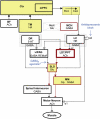
Schematic illustrating the brain regions currently known to be involved in the control of wakefulness and muscle tone. Neuronal populations that are active during wakefulness (green) consist of hypocretin neurons (Hcrt) that project most densely and provide excitatory input (solid arrowheads) to the locus coeruleus (LC) and other wake-promoting areas: the basal forebrain (BF), tuberomamillary nucleus (TM), dorsal raphe (DR), laterodorsal and pedunculopontine tegmental nuclei (LDT/PPT), ventrolateral periaqueductal gray (vlPAG), and directly to cortex and spinal motor neurons. Hcrt neurons also increase motor tone through suppression of REM sleep atonia circuitry, which consists of sublaterodorsal nucleus (SLD) excitation of medial medulla (MM) and spinal interneurons that inhibit motor neurons. Active inhibition of atonia circuitry is mediated by HCRT excitation of monoaminergic and GABAergic pathways that inhibit (blunt terminals) the SLD. Disinhibition of REM sleep atonia circuitry during wakefulness may underlie cataplexy in narcolepsy (see Fig. 4). Key: solid lines/arrows, active excitation; solid lines/blunt terminals, active inhibition; dashed lines/arrows, disfacilitation; dashed lines/blunt terminals, disinhibition; thick black lines, Hcrt projections; red, neurons and pathways that are active in REM sleep; REMoff, neurons that are silent in REM sleep; Ctx, Cerebral cortex; mPFC, medial prefrontal cortex; BLA, basolateral amygdala; CeA, central nucleus of the amygdala; MCH, melanin-concentrating hormone cells; ACh, acetylcholine; HA, histamine; Glu, glutamate; GABA, gamma-aminobutyric acid; Gly, glycine; 5-HT, serotonin; NE, norepinephrine; α2, noradrenergic autoreceptor; α1, noradrenergic α1 receptor.
Fig. 4
Fig. 4
Schematic illustrating arousal-state circuitry in narcolepsy and cataplexy. Hypocretin (Hcrt) neurons and their projections to wake-promoting regions (see Fig. 3) are absent in narcolepsy. Presumably as a compensatory consequence, pontine cholinergic supersensitivity (thick lined box), monoaminergic hypoactivity in the amygdala (thin lined boxes) and increased numbers of HA neurons develop. Neuronal populations that are active during cataplexy (yellow) include regions that are also active during wakefulness and REM sleep. Cataplexy can be triggered by positive emotional stimuli that activate neurons in the mPFC and amygdala, which then may disinhibit REM sleep atonia circuitry. In individuals without narcolepsy, HCRT excitation onto LC neurons could balance the GABAergic inhibition from CeA to maintain normal muscle tone. Activation of the atonia circuitry is mediated by withdrawal of GABAergic inhibition of SLD neurons from the vlPAG and monoaminergic inhibition from the DR and LC. Direct activation of SLD neurons could result from upregulated cholinergic mechanisms in the LDT/PPT. Cataplexy can be alleviated by antidepressants that increase noradrenergic tone via blockade of α2 autoreceptors; mechanisms of GABAB therapeutics for cataplexy are unknown, but may involve inhibition of SLD neurons to suppress atonia. Key: see Fig. 3.
Fig. 5
Fig. 5
The narcolepsy/cataplexy assay for orexin/ataxin-3 transgenic mice. (A) Cataplexy density (the number of cataplexy bouts per time awake) and (B) percent time awake during the 6 h following pretreatment with almorexant (ALM, black) vs. vehicle (VEH, white) at ZT12 and treatment with desipramine (DES 0–5 mg/kg) 30 min later. Two-way repeated measures ANOVA: * p < 0.05 vs. VEH pretreatment, *p < 0.05 vs. DES (0 mg/kg); n = 4.

References

    1. Absalom N, Eghorn LF, Villumsen IS, Karim N, Bay T, Olsen JV, Knudsen GM, Brauner-Osborne H, Frolund B, Clausen RP, Chebib M, Wellendorph P. alpha4betadelta GABAA receptors are high-affinity targets for gamma-hydroxybutyric acid (GHB). Proc. Natl. Acad. Sci. U.S.A. 2012;109:13404–13409. - PMC - PubMed
    1. Ahmed SS, Schur PH, MacDonald NE, Steinman L. Narcolepsy, 2009 A(H1N1) pandemic influenza, and pandemic influenza vaccinations: what is known and unknown about the neurological disorder, the role for autoimmunity, and vaccine adjuvants. J. Autoimmun. 2014;50:1–11. - PubMed
    1. Ahmed SS, Volkmuth W, Duca J, Corti L, Pallaoro M, Pezzicoli A, Karle A, Rigat F, Rappuoli R, Narasimhan V, Julkunen I, Vuorela A, Vaarala O, Nohynek H, Pasini FL, Montomoli E, Trombetta C, Adams CM, Rothbard J, Steinman L. Antibodies to influenza nucleoprotein cross-react with human hypocretin receptor 2. Sci. Transl. Med. 2015;7:294ra105. - PubMed
    1. Akimoto H, Honda Y, Takahashi Y. Pharmacotherapy in narcolepsy. Dis. Nerv. Syst. 1960;21:704–706. - PubMed
    1. Alexander SP, Benson HE, Faccenda E, Pawson AJ, Sharman JL, Spedding M, Peters JA, Harmar AJ, Collaborators C. The Concise Guide to PHARMACOLOGY 2013/14: G protein-coupled receptors. Br. J. Pharmacol. 2013;170:1459–1581. - PMC - PubMed

Publication types