Review
. 2016 Jan;68(1):142-67.
doi: 10.1124/pr.114.010090.

Toll-like Receptors in the Vascular System: Sensing the Dangers Within

Affiliations
Review

Toll-like Receptors in the Vascular System: Sensing the Dangers Within

Styliani Goulopoulou et al. Pharmacol Rev. 2016 Jan.

Abstract

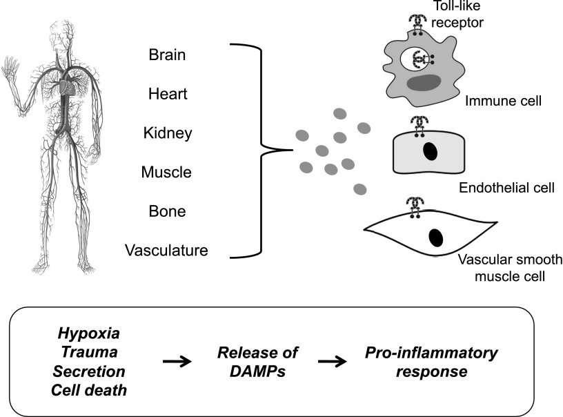
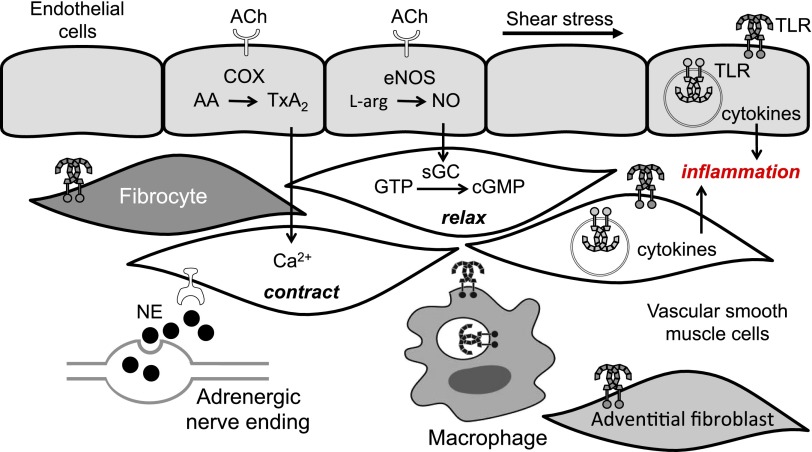
Toll-like receptors (TLRs) are components of the innate immune system that respond to exogenous infectious ligands (pathogen-associated molecular patterns, PAMPs) and endogenous molecules that are released during host tissue injury/death (damage-associated molecular patterns, DAMPs). Interaction of TLRs with their ligands leads to activation of downstream signaling pathways that induce an immune response by producing inflammatory cytokines, type I interferons (IFN), and other inflammatory mediators. TLR activation affects vascular function and remodeling, and these molecular events prime antigen-specific adaptive immune responses. Despite the presence of TLRs in vascular cells, the exact mechanisms whereby TLR signaling affects the function of vascular tissues are largely unknown. Cardiovascular diseases are considered chronic inflammatory conditions, and accumulating data show that TLRs and the innate immune system play a determinant role in the initiation and development of cardiovascular diseases. This evidence unfolds a possibility that targeting TLRs and the innate immune system may be a novel therapeutic goal for these conditions. TLR inhibitors and agonists are already in clinical trials for inflammatory conditions such as asthma, cancer, and autoimmune diseases, but their study in the context of cardiovascular diseases is in its infancy. In this article, we review the current knowledge of TLR signaling in the cardiovascular system with an emphasis on atherosclerosis, hypertension, and cerebrovascular injury. Furthermore, we address the therapeutic potential of TLR as pharmacological targets in cardiovascular disease and consider intriguing research questions for future study.


Figures

Fig. 1.
Fig. 1.
Cell surface and intracellular Toll-like receptors (TLRs) and their ligands. TLRs are divided into two groups based on their cellular localization when sensing their respective ligands. TLRs 1, 2, 4–6, and 11 localize to the cell surface (cell surface TLRs) and TLRs 3 and 7–9 reside at endosomal compartments (intracellular TLRs). Cell surface TLRs respond to microbial membrane materials such as lipids, lipoproteins, and proteins, whereas intracellular TLRs recognize bacteria- and virus-derived nucleic acids.
Fig. 2.
Fig. 2.
Exogenous and endogenous Toll-like receptor ligands. Categories of pathogen-associated molecular patterns (exogenous ligands) and damage-associated molecular patterns (endogenous ligands). HMBG1, high-mobility group box 1; HSP, heat shock protein; LDL, low-density lipoprotein; LPS, lipopolysaccharide; MRP, myeloid related protein; TFAM, mitochondrial transcription factor A
Fig. 3.
Fig. 3.
Toll-like receptors, adapter proteins, and signaling molecules. With the exception of TLR3, all TLRs recruit the adapter protein, myeloid differentiation primary response gene 88 (MyD88). In addition, TLRs 1, 2, 4, and 6 recruit the adapters cluster of differentiation 14 (CD14, not shown), which is required for lipopolysaccharide (LPS) binding, and Toll-Interleukin 1 receptor domain containing adaptor protein (TIRAP), which links the conserved C-terminal intracellular Toll/interleukin-1 receptor (TIR) domain with MyD88. In the MyD88-dependent pathway, the MyD88 recruits interleukin 1 receptor-associated kinase (IRAK), which interacts with the adapter protein tumor necrosis factor-receptor-associated factor 6 (TRAF6) and provides a link to nuclear factor kappa-light-chain-enhancer of activated B cells (NF-κB) translocation. The MyD88-dependent pathway also facilitates expression of mitogen-activated protein kinase (MAPK) and transcription factors, such as interferon regulatory factors (IRFs), driving the production of proinflammatory mediators, including cytokines and chemokines. Activation of TLR3 initiates the TIR-domain-containing adapter-inducing interferon-β (TRIF)-dependent pathway, whereas TLR4 can signal via either MyD88-dependent or TRIF-dependent pathways requiring the additional linker adaptor TRIF-related adaptor molecule (TRAM) to associate with TRIF. In the TRIF-dependent pathway, TRIF interacts with TRAF3 to activate IRF3 initiating IFN-β production, which is the hallmark of the host innate response to viral infection.
Fig. 4.
Fig. 4.
Damage-associated molecular pattern (DAMP)-induced activation of Toll-like receptors (TLRs). Schematic demonstrating the concept that circulating damage-associated molecular patterns (DAMPs) released after hypoxia, trauma, and cell death lead to TLR activation in immune cells, endothelial cells, and vascular smooth muscle cells. Prolonged or excessive activation of TLRs on these cells provides a proinflammatory state, leading to endothelial dysfunction and subsequent cardiovascular disease.
Fig. 5.
Fig. 5.
Toll-like receptor (TLR) localization in cellular components of the vascular wall. TLR activation by damage-associated molecular patterns (DAMPs) sets an inflammatory response in the vessel wall and interferes with endothelium-dependent relaxation and vasoconstriction. TLR activation on endothelial cells contributes to decreased production and bioavailability of nitric oxide, exacerbated formation of cytokines and reactive oxygen species, and the release of vasoconstrictor metabolites of arachidonic acid. TLR activation on smooth muscle cells increases formation of reactive oxygen species and reduces calcium sequestration in the sarcoplasmic reticulum. Adventitial fibroblasts and fibrocytes are newly identified components of the vascular wall that may contribute to proinflammatory events via activation of TLRs. AA, arachidonic acid; ACh, acetylcholine; cGMP, cyclic guanosine monophosphate; COX, cyclooxygenase; eNOS, endothelial nitric oxide synthase; GTP, guanosine-5′-triphosphate; l-arg, l-arginine; NE, norepinephrine; NO, nitric oxide; sGC, soluble guanylyl cyclase; TxA2; thromboxane A2. Revised image based on a previously published Figure from FASEB J; reproduced by permission.

References

    1. Adachi N, Hirota M, Hamaguchi M, Okamoto K, Watanabe K, Endo F. (2004) Serum cytochrome c level as a prognostic indicator in patients with systemic inflammatory response syndrome. Clin Chim Acta 342:127–136. - PubMed
    1. Akira S, Uematsu S, Takeuchi O. (2006) Pathogen recognition and innate immunity. Cell 124:783–801. - PubMed
    1. Alexopoulou L, Holt AC, Medzhitov R, Flavell RA. (2001) Recognition of double-stranded RNA and activation of NF-kappaB by Toll-like receptor 3. Nature 413:732–738. - PubMed
    1. An SJ, Liu P, Shao TM, Wang ZJ, Lu HG, Jiao Z, Li X, Fu JQ. (2015) Characterization and functions of vascular adventitial fibroblast subpopulations. Cell Physiol Biochem 35:1137–1150. - PubMed
    1. Anderson KV, Jürgens G, Nüsslein-Volhard C. (1985) Establishment of dorsal-ventral polarity in the Drosophila embryo: genetic studies on the role of the Toll gene product. Cell 42:779–789. - PubMed

Publication types

MeSH terms

Substances

LinkOut - more resources