. 2016 Feb 19;291(8):4247-55.
doi: 10.1074/jbc.M115.697888. Epub 2015 Dec 31.

CXCL1/MGSA Is a Novel Glycosaminoglycan (GAG)-binding Chemokine: STRUCTURAL EVIDENCE FOR TWO DISTINCT NON-OVERLAPPING BINDING DOMAINS

Affiliations

CXCL1/MGSA Is a Novel Glycosaminoglycan (GAG)-binding Chemokine: STRUCTURAL EVIDENCE FOR TWO DISTINCT NON-OVERLAPPING BINDING DOMAINS

Krishna Mohan Sepuru et al. J Biol Chem. .

Abstract

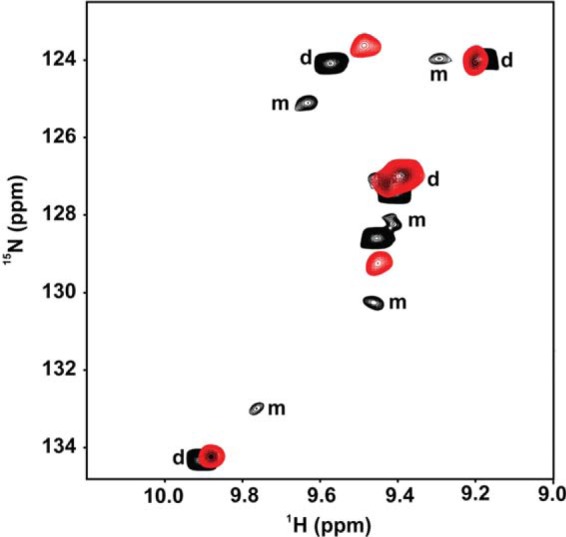
In humans, the chemokine CXCL1/MGSA (hCXCL1) plays fundamental and diverse roles in pathophysiology, from microbial killing to cancer progression, by orchestrating the directed migration of immune and non-immune cells. Cellular trafficking is highly regulated and requires concentration gradients that are achieved by interactions with sulfated glycosaminoglycans (GAGs). However, very little is known regarding the structural basis underlying hCXCL1-GAG interactions. We addressed this by characterizing the binding of GAG heparin oligosaccharides to hCXCL1 using NMR spectroscopy. Binding experiments under conditions at which hCXCL1 exists as monomers and dimers indicate that the dimer is the high-affinity GAG ligand. NMR experiments and modeling studies indicate that lysine and arginine residues mediate binding and that they are located in two non-overlapping domains. One domain, consisting of N-loop and C-helical residues (defined as α-domain) has also been identified previously as the GAG-binding domain for the related chemokine CXCL8/IL-8. The second domain, consisting of residues from the N terminus, 40s turn, and third β-strand (defined as β-domain) is novel. Eliminating β-domain binding by mutagenesis does not perturb α-domain binding, indicating two independent GAG-binding sites. It is known that N-loop and N-terminal residues mediate receptor activation, and we show that these residues are also involved in extensive GAG interactions. We also show that the GAG-bound hCXCL1 completely occlude receptor binding. We conclude that hCXCL1-GAG interactions provide stringent control over regulating chemokine levels and receptor accessibility and activation, and that chemotactic gradients mediate cellular trafficking to the target site.

Keywords: CXC chemokines; CXCL1/MGSA; G protein-coupled receptor (GPCR); NMR; cell migration; chemokine; glycobiology; glycosaminoglycan; heparin; immunology.


Figures

FIGURE 1.
FIGURE 1.
Ribbon representation of a WT hCXCL1 dimer and single monomeric unit of the dimer structures (PDB code 1MGS). A and B, the individual monomers in the dimer are shown in dark and light blue for clarity.
FIGURE 2.
FIGURE 2.
The hCXCL1 dimer is the high-affinity GAG ligand. A section of the 1H,15N HSQC spectra showing the overlay of WT hCXCL1 in the free (black) and GAG-bound (red) forms. Dimer (d) and monomer (m) peaks are indicated. The monomer peaks disappear on dp14 binding, indicating that the dimer is the high-affinity GAG ligand. The spectra were collected using a 15 μm hCXCL1 sample in 50 mm sodium phosphate (pH 6.0) at 40 °C.
FIGURE 3.
FIGURE 3.
Binding of WT hCXCL1 to heparin GAGs. A, sections of the 1H,15N HSQC spectra showing the overlay of WT hCXCL1 in the free (black) and dp14-bound (red) forms. Arrows indicate the direction of the peak movement. B and C, histograms of chemical shift changes in the hCXCL1 dimer on binding heparin dp8 (B) and dp14 (C). The basic residues Lys, Arg, and His are shown in blue, and buried residues (ASA < 20%) are shown in black. The CSP of Lys-21 is truncated, and the actual CSP is 1.80 ppm. The horizontal line at 0.1 ppm represents the cutoff for a residue to be considered perturbed. The spectra were collected using a 100 μm hCXCL1 sample in 50 mm sodium phosphate (pH 5.7) at 40 °C.
FIGURE 4.
FIGURE 4.
Backbone dynamics of the hCXCL1-GAG complex. Comparison of 15N,1H NOE values for hCXCL1 in the free (black) and dp14-bound (red) forms. The data show that the N-terminal, N-loop, and 30s loop residues in the GAG-bound form have higher NOE values. A NOE difference plot between the bound and free forms (NOEbound − NOEfree) is shown as an inset. The spectra were collected using a 100 μm hCXCL1 sample in 50 mm sodium phosphate (pH 5.7) at 40 °C.
FIGURE 5.
FIGURE 5.
Binding of the hCXCL1 R8A mutant to heparin GAG. Shown is a histogram of chemical shift changes in the hCXCL1 R8A mutant on binding heparin dp14. The data indicate binding only to the α-domain. The basic residues Lys, Arg, and His are shown in blue, and buried residues (ASA < 20%) are shown in black. The CSP of Lys-21 is truncated, and the actual CSP is 1.85 ppm. The horizontal line at 0.1 ppm represents the cutoff for a residue to be considered perturbed. The spectra were collected using a 100 μm hCXCL1 sample in 50 mm sodium phosphate (pH 5.7) at 40 °C.
FIGURE 6.
FIGURE 6.
Binding of hCXCL1 to the CXCR2 N-domain. Shown is a histogram of chemical shift changes in WT hCXCL1 on binding the CXCR2 N-domain peptide. The data show that the N-loop and the adjacent third β-strand residues mediate binding. The basic residues Lys, Arg, and His are shown in blue, and buried residues (ASA < 20%) are shown in black. The horizontal line at 0.07 ppm represents the cutoff for a residue to be considered perturbed. The spectra were collected using a 100 μm hCXCL1 sample in 50 mm sodium phosphate (pH 5.7) at 40 °C.
FIGURE 7.
FIGURE 7.
GAG-bound hCXCL1 cannot bind the receptor. A, sections of the 1H,15N HSQC spectra showing the overlay of several residues in the free form (black), the GAG-bound form (blue, left panel), the CXCR2 N-domain-bound form (red, center panel), and on titrating the CXCR2 N-domain peptide to the GAG-bound hCXCL1 (magenta, right panel). B, surface representation showing GAG binding (blue), receptor binding (red), and shared binding (yellow) regions. Residues that are solvent-exposed but occluded in the GAG-bound form (ASA <20%) were considered to be involved in binding. For receptor binding, residues that showed significant CSP and were solvent-exposed and N-terminal Glu-6 and Leu-7 (of the ELR motif) were considered to be involved in binding.
FIGURE 8.
FIGURE 8.
GAG binding domains in hCXCL1. A–D, surface plots highlighting GAG binding to the α-domain (A and C) and β-domain (B and D). A and B, the hCXCL1 dimer is shown in ribbon representation, and GAG is shown as sticks. C and D, the geometry of the GAG chain and interactions of the GAG-binding residues. In the two monomers, GAG-binding residues are shown in light and dark blue, respectively. E, GAG can bind both domains without steric clashes. Two GAG chains are represented as spheres. We used the hCXCL1 dimer (PDB code 1MGS) and heparin 12-mer (PDB code 1HPN) structures to generate the HADDOCK models.
FIGURE 9.
FIGURE 9.
Sequences of ELR chemokines. Basic residues of the α-domain (red) and β-domain (blue) in CXCL1 and the corresponding regions in other members are highlighted.
FIGURE 10.
FIGURE 10.
Models of ELR chemokine-heparan sulfate interactions. A, proposed clamp model showing that hCXCL1 is sandwiched between the NS domains of heparan sulfate. B, proposed horseshoe model for the CXCL8-heparan sulfate complex (33). C, schematic showing binding of two adjacent proteoglycan (PG) heparan sulfate chains to the hCXCL1 dimer.

References

    1. Bonecchi R., Galliera E., Borroni E. M., Corsi M. M., Locati M., and Mantovani A. (2009) Chemokines and chemokine receptors: an overview. Front. Biosci. 14, 540–551 - PubMed
    1. Griffith J. W., Sokol C. L., and Luster A. D. (2014) Chemokines and chemokine receptors: positioning cells for host defense and immunity. Annu. Rev. Immunol. 32, 659–702 - PubMed
    1. Das S. T., Rajagopalan L., Guerrero-Plata A., Sai J., Richmond A., Garofalo R. P., and Rajarathnam K. (2010) Monomeric and dimeric CXCL8 are both essential for in vivo neutrophil recruitment. PLoS ONE 5, e11754. - PMC - PubMed
    1. Gangavarapu P., Rajagopalan L., Kolli D., Guerrero-Plata A., Garofalo R. P., and Rajarathnam K. (2012) The monomer-dimer equilibrium and glycosaminoglycan interactions of chemokine CXCL8 regulate tissue-specific neutrophil recruitment. J. Leukocyte Biol. 91, 259–265 - PMC - PubMed
    1. Li L., Ly M., and Linhardt R. J. (2012) Proteoglycan sequence. Mol. Biosyst. 8, 1613–1625 - PMC - PubMed

Publication types

LinkOut - more resources