. 2016 Mar 1;25(5):892-902.
doi: 10.1093/hmg/ddv618. Epub 2015 Dec 31.

TRIO loss of function is associated with mild intellectual disability and affects dendritic branching and synapse function

Affiliations

TRIO loss of function is associated with mild intellectual disability and affects dendritic branching and synapse function

Wei Ba et al. Hum Mol Genet. .

Abstract

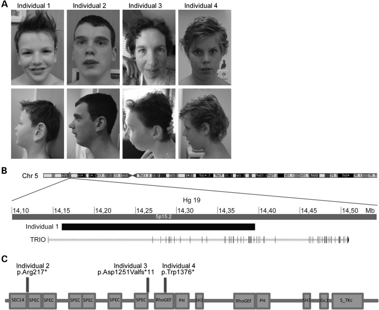
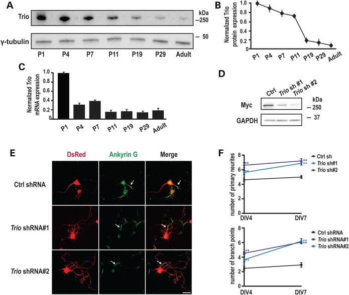
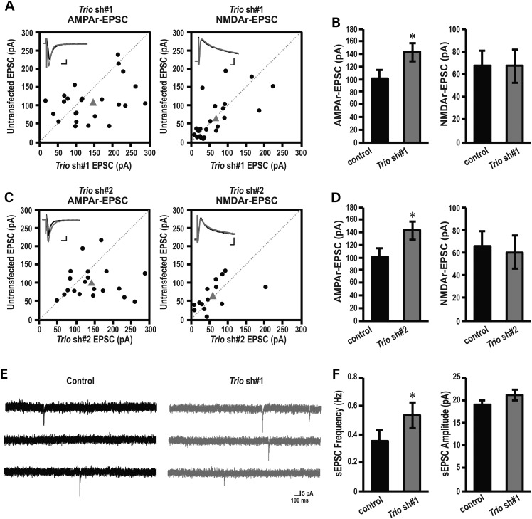
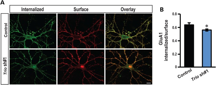
Recently, we marked TRIO for the first time as a candidate gene for intellectual disability (ID). Across diverse vertebrate species, TRIO is a well-conserved Rho GTPase regulator that is highly expressed in the developing brain. However, little is known about the specific events regulated by TRIO during brain development and its clinical impact in humans when mutated. Routine clinical diagnostic testing identified an intragenic de novo deletion of TRIO in a boy with ID. Targeted sequencing of this gene in over 2300 individuals with ID, identified three additional truncating mutations. All index cases had mild to borderline ID combined with behavioral problems consisting of autistic, hyperactive and/or aggressive behavior. Studies in dissociated rat hippocampal neurons demonstrated the enhancement of dendritic formation by suppressing endogenous TRIO, and similarly decreasing endogenous TRIO in organotypic hippocampal brain slices significantly increased synaptic strength by increasing functional synapses. Together, our findings provide new mechanistic insight into how genetic deficits in TRIO can lead to early neuronal network formation by directly affecting both neurite outgrowth and synapse development.


Figures

Figure 1.
Figure 1.
Individuals with LoF TRIO mutations. (A) Frontal and lateral photographs of Individual 1 with the deletion disrupting TRIO and Individuals 2–4 with LoF mutations in TRIO. Only mild facial dysmorphisms were observed. (B) Schematic overview of the 235 kb de novo deletion on chromosome 5, partially disrupting TRIO in Individual 1. (C) Schematic overview of TRIO, including the known domains (N-terminal SEC14 domain, several spectrin repeats, two DH-PH Rho-GEF units, Ig-like domain and C-terminal putative serine/threonine kinase domain). The positions of the three identified mutations in Individuals 2–4 (p.Trp1376*, p.Asp1251Valfs*11 and p.(Arg217*) are depicted.
Figure 2.
Figure 2.
Developmental expression of TRIO and its role in neurite outgrowth. (A) Rat hippocampi were collected at the indicated ages; equal amounts of protein (50 μg) were subjected to western blot analysis and representative blots are shown. (B) Quantification of Trio protein levels at different postnatal ages, normalized to the levels at P1 (n = 3). (C) Quantification of Trio mRNA levels using Q-PCR at different postnatal ages (n = 3). (D) 293 cells co-transfected with vectors encoding MYC-TRIO and Trio shRNA 1 and 2 were harvested after 24 h. Western blot analysis using a MYC antibody demonstrated that both Trio shRNA 1 and 2 efficiently reduced Trio expression. (E) Rat hippocampal neurons nucleofected at the time of plating with a control (scrambled) shRNA or Trio shRNA 1 or 2 were fixed and analyzed at the indicated times. shRNA transfected neurons were identified by DsRed co-expression; axon initial segments were identified by staining for Ankyrin G; representative images at DIV7 are shown. Scale bar, 50 µm. (F) Quantification of primary neurites and branch points at DIV4 and DIV7. Neurons expressing Trio shRNAs exhibited more primary neurites and more branch points than neurons expressing the control shRNA. Five separate experiments were performed; 20–30 neurons per group per experiment were counted. **P < 0.01 with respect to the control (scramble) shRNA vector.
Figure 3.
Figure 3.
Post-synaptic TRIO regulates synaptic transmission. (A, B) Amplitudes of AMPAr (left panel) and NMDAr (right panel) eEPSCs in control, non-transfected neurons are plotted against simultaneously recorded neighboring neurons expressing (A) Trio shRNA 1 (AMPAr, n = 25; NMDAr, n = 22), (B) Trio shRNA 2 (AMPAr, n = 20; NMDAr, n = 19). Black symbols represent single pairs of recordings; grey symbols show mean amplitudes. Inserts in each panel show sample averaged traces; grey traces, transfected neurons; black traces, non-transfected neighboring neurons. Scale bars represent 10 ms and 25 pA. (C and D) Summary (mean ± SEM) of effects of expressing Trio shRNA 1, on AMPAr (left) and NMDAr (right) eEPSCs calculated as the averaged ratios obtained from pairs of infected and uninfected neighboring neurons. Data are shown as mean ± SEM. *P < 0.05, paired Student's t-test, Trio shRNA 1 (AMPAr, n = 25; NMDAr, n = 22), (B) Trio shRNA 2 (AMPAr, n = 20; NMDAr, n = 19). (E) Representative traces of sEPSC recorded at −60 mV in an individual neuron from untransfected (control) or Trio shRNA 1 transfected group. Scale bars represent 100 ms and 5 pA. (F) Quantification of sEPSC frequency (left) and amplitude (right) for control and Trio shRNA 1-expressing neurons. Data are shown as mean ± SEM. *P < 0.05, paired Student's t-test, n = 8 pairs.
Figure 4.
Figure 4.
Down-regulation of Trio reduces AMPAr endocytosis. (A) Representative double-label images of internalized (green) and surface (red) AMPAr GluA1 subunit in low-density hippocampal neurons. (B) Ratiometric analysis of the intensity of internalized GluA1 to surface GluA1 in indicated conditions. Control shRNA: n = 15; Trio shRNA 1: n = 14; scale bars, 10 µm; *P < 0.05, t-test; error bars represent SEM.

References

    1. Leonard H., Wen X. (2002) The epidemiology of mental retardation: challenges and opportunities in the new millennium. Ment. Retard. Dev. Disabil. Res. Rev., 8, 117–134. - PubMed
    1. Vissers L.E.L.M., Gilissen C., Veltman J.A. (2015) Genetic studies in intellectual disability and related disorders. Nat. Rev. Genet. 10.1038/nrg3999. - DOI - PubMed
    1. Bamshad M.J., Ng S.B., Bigham A.W., Tabor H.K., Emond M.J., Nickerson D.A., Shendure J. (2011) Exome sequencing as a tool for Mendelian disease gene discovery. Nat. Rev. Genet., 12, 745–755. - PubMed
    1. Yang Y., Muzny D.M., Reid J.G., Bainbridge M.N., Willis A., Ward P.A., Braxton A., Beuten J., Xia F., Niu Z. et al. (2013) Clinical whole-exome sequencing for the diagnosis of Mendelian disorders. N. Engl. J. Med., 369, 1502–1511. - PMC - PubMed
    1. Vissers L.E.L.M., de Ligt J., Gilissen C., Janssen I., Steehouwer M., de Vries P., van Lier B., Arts P., Wieskamp N., del Rosario M. et al. (2010) A de novo paradigm for mental retardation. Nat. Genet., 42, 1109–1112. - PubMed

Publication types

MeSH terms

Substances