. 2016 Jan 1;351(6268):88-91.
doi: 10.1126/science.aad4992.

Structure of the Sec61 channel opened by a signal sequence

Affiliations

Structure of the Sec61 channel opened by a signal sequence

Rebecca M Voorhees et al. Science. .

Abstract

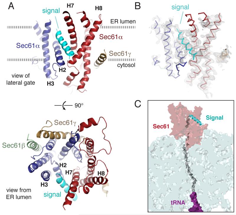
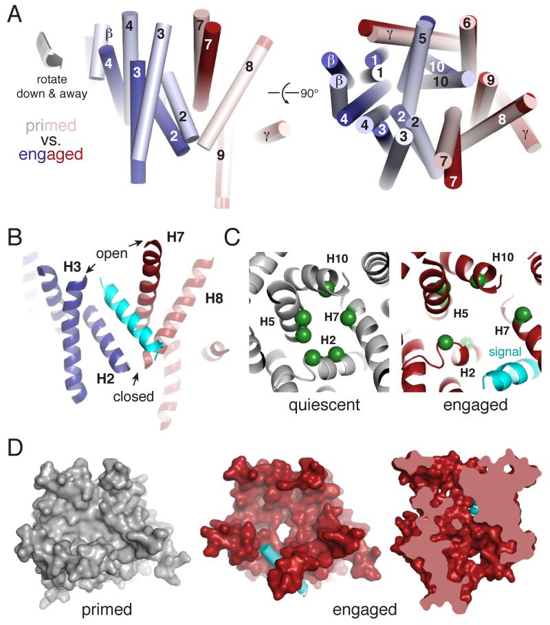
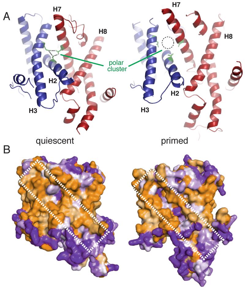
Secreted and integral membrane proteins compose up to one-third of the biological proteome. These proteins contain hydrophobic signals that direct their translocation across or insertion into the lipid bilayer by the Sec61 protein-conducting channel. The molecular basis of how hydrophobic signals within a nascent polypeptide trigger channel opening is not understood. Here, we used cryo-electron microscopy to determine the structure of an active Sec61 channel that has been opened by a signal sequence. The signal supplants helix 2 of Sec61α, which triggers a rotation that opens the central pore both axially across the membrane and laterally toward the lipid bilayer. Comparisons with structures of Sec61 in other states suggest a pathway for how hydrophobic signals engage the channel to gain access to the lipid bilayer.


Figures

Fig. 1
Fig. 1. Structure of the signal peptide-engaged Sec61 complex
(A) View of the lateral gate (top) or from the ER lumen of the Sec61 complex bound to the pre-prolactin signal peptide (cyan). The mobile regions of Sec61α are blue, while the comparatively immobile regions are red. The β and γ subunits are pale green and tan, respectively. Helices that comprise the lateral gate are labelled. (B) Experimental density for the structure (mesh, filtered to 4.5 Å resolution) superimposed on the backbone trace of the structural model. (C) Density observed for the nascent polypeptide through the ribosomal tunnel and parts of the Sec61channel.
Fig. 2
Fig. 2. Conformational changes to Sec61 upon engagement by a signal peptide
(A) Positions of the transmembrane helices of the Sec61 complex in the ribosome-primed (pale colors, PDB 3J7Q) and signal-engaged (bright colors) states. The mobile regions of Sec61α are blue, while the comparatively immobile regions are red. Individual helices are labelled; the signal has been omitted for clarity. (B) View of the asymmetrically opened lateral gate. (C) View of the pore ring residue positions (green spheres) in the quiescent SecY crystal structure (grey, PDB 1RH5) and engaged Sec61 complex (red). The signal is cyan. (D) Left and middle: surface view of the primed (grey, PDB 3J7Q) and engaged (red) states of the Sec61 complex viewed from the ER lumen. The lumenal loops have been removed for clarity. The signal peptide is cyan. Right: cutaway view perpendicular to the membrane of the engaged Sec61 complex showing an open channel.
Fig. 3
Fig. 3. The lateral gate is destabilized in the ribosome-primed Sec61 complex
(A) Comparison of the quiescent SecY (left, PDB 1RH5) and primed Sec61 (right, PDB 3J7Q) complexes. Ribosome binding results in partial destabilization of the lateral gate by shifting helices 2 and 3 away from the midline, which disrupts the polar cluster (green). (B) Space filling model viewing the lateral gate of the quiescent (left) and primed (right) structures colored by hydrophobicity, in which orange is hydrophobic and purple is hydrophilic. The hydrophilic seam produced by ribosome binding is indicated.
Fig. 4
Fig. 4. Putative path of the signal peptide into the Sec61 lateral gate
(A) Space-filling model of the cytosolic vestibule of Sec61 viewed from the ribosome, colored by hydrophobicity as in Fig. 3B. (B) Residues comprising the hydrophobic patch are at the lateral gate. (C) The eventual position of the signal peptide (cyan), relative to the stationary ribosome-bound regions of Sec61, is essentially identical to the position of helix 2 in the quiescent state (grey). (D) Positions of helix 2 in the quiescent (grey) and primed (yellow) states superimposed on the engaged Sec61 structure (red). The signal peptide is omitted for clarity, but would reside precisely in the position of the quiescent helix 2.

References

    1. Park E, Rapoport TA. Mechanisms of Sec61/SecY-mediated protein translocation across membranes. Annu Rev Biophys. 2012;41:21–40. - PubMed
    1. Van den Berg B, et al. X-ray structure of a protein-conducting channel. Nature. 2004;427:36–44. - PubMed
    1. Plath K, Mothes W, Wilkinson BM, Stirling CJ, Rapoport TA. Signal sequence recognition in posttranslational protein transport across the yeast ER membrane. Cell. 1998;94:795–807. - PubMed
    1. Egea PF, Stroud RM. Lateral opening of a translocon upon entry of protein suggests the mechanism of insertion into membranes. Proc. Natl. Acad. Sci. U.S.A. 2010;107:17182–17187. - PMC - PubMed
    1. Tsukazaki T, et al. Conformational transition of Sec machinery inferred from bacterial SecYE structures. Nature. 2008;455:988–991. - PMC - PubMed

Publication types