. 2016 Jan;36(1):49-60.

A Novel Sirtuin-3 Inhibitor, LC-0296, Inhibits Cell Survival and Proliferation, and Promotes Apoptosis of Head and Neck Cancer Cells

Affiliations

A Novel Sirtuin-3 Inhibitor, LC-0296, Inhibits Cell Survival and Proliferation, and Promotes Apoptosis of Head and Neck Cancer Cells

Turki Y Alhazzazi et al. Anticancer Res. 2016 Jan.

Abstract

Background: The survival rate of patients with head and neck squamous cell carcinoma (HNSCC) stands at approximately 50% and this has not improved in decades. This study developed a novel sirtuin-3 (SIRT3) inhibitor (LC-0296) and examined its role in altering HNSCC tumorigenesis.

Materials and methods: The effect of the SIRT3 inhibitor, LC-0296, on cell survival, proliferation, and apoptosis, and reactive oxygen species levels in HNSCC cells were studied.

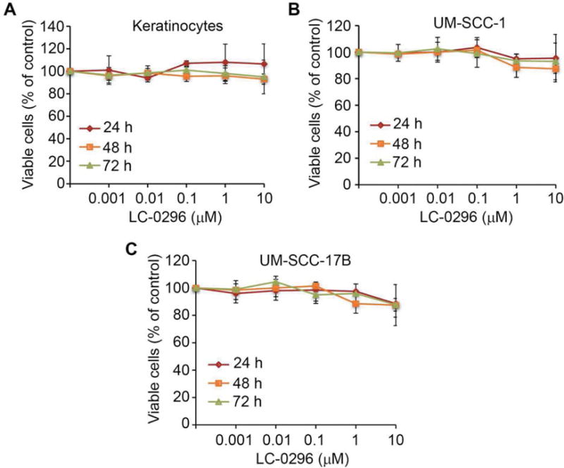
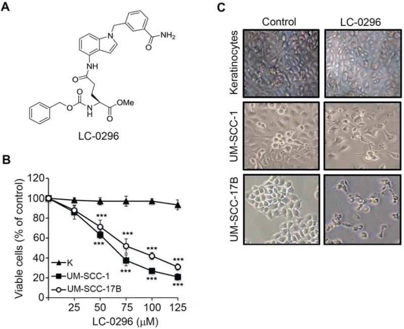
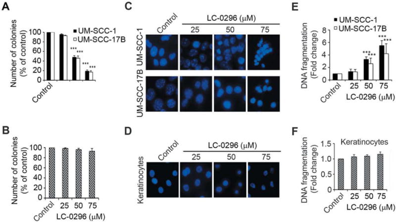
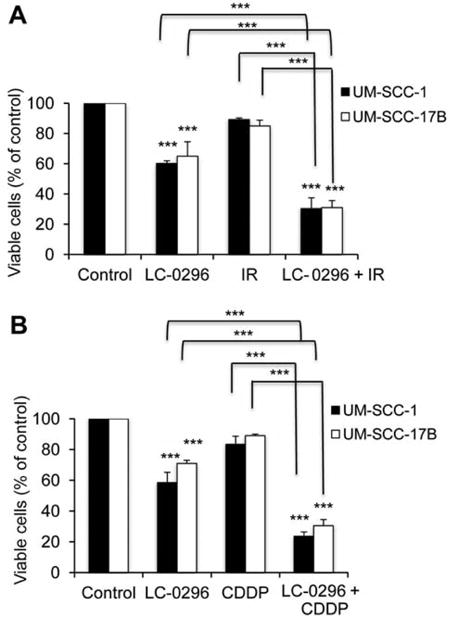
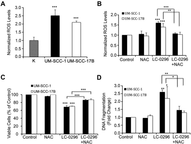
Results: LC-0296 reduces cell proliferation and promotes apoptosis of HNSCC cells but not of normal human oral keratinocytes. This inhibitory effect is mediated, in part, via modulation of reactive oxygen species levels. Additionally, LC-0296 works synergistically to increase the sensitivity of HNSCC cells to radiation and cisplatin treatment.

Conclusion: Development of novel SIRT3 inhibitors, such as LC-0296, might enable the development of new targeted therapies to treat and improve the survival rate of patients with head and neck cancer.

Keywords: HNSCC; ROS; SIRT3; Sirtuins; oral cancer; sirtuin inhibitor; sirtuin-3.


Figures

Figure 1
Figure 1
A: Schematic chemical synthesis of the sirtuin-3 (SIRT3) inhibitor (LC-0296). Commercially available 4-nitro-1H-indole (1) was alkylated to give compound 3, whose nitro group was reduced to the corresponding amine in compound 4 in excellent yields. The methyl ester group in compound 4 was converted into a primary amide with methanolic ammonia. The resulting compound 5 was coupled with L-glutamate derived Z-Glu-OMe in the presence of 1-ethyl-3-(3-dimethylaminopropyl) carbodiimide hydrochloride (EDC) and 1-hydroxybenzotriazole (HOBt) to yield LC-0296 (6) in good yields. Reaction conditions were: (a) NaH, DMF, yield 68%; (b) NiCl2·6H2O, NaBH4, MeOH, yield 89%; (c) methanolic NH3, CaCl2, 70°C, yield 85%; (d) Z-Glu-OMe, EDC, HOBt, NMM, CH2Cl2, yield 60%. B: Effect of the sirtuin-3 (SIRT3) inhibitor, LC-0296 on sirtuin-1 (SIRT1), sirtuin-2 (SIRT2) and sirtuin-3 (SIRT3) in an in vitro enzymatic assay.
Figure 2
Figure 2
The effect of the sirtuin-3 (SIRT3) inhibitor, LC-0296, on head and neck squamous cell carcinoma cells and keratinocytes using low doses. Normal human oral keratinocytes (A), UM-SCC-1 (B), and UM-SCC-17B (C) cells were seeded in 96-well plates at 5×10³ cells/well, then treated with dimethyl sulfoxide (control) or LC-0296, as indicated for 24, 48, and 72 h. Cell viability was then determined by the QUANT Cell Proliferation Assay Kit. The graphs illustrate the percentage of viable cells following treatment.
Figure 3
Figure 3
Sirtuin-3 (SIRT3) inhibitor, LC-0296, inhibits head and neck squamous cell carcinoma (HNSCC) cell survival without affecting normal human oral keratinocytes. A: The chemical structure of the SIRT3 inhibitor LC-0296 is shown. B: HNSCC cells (UM-SCC-1 and UM-SCC-17B) and normal human oral keratinocytes (K) were seeded in 96-well plates at 5×10³ cells/well then treated with LC-0296 as indicated for 24 h. Cell viability was determined by the QUANT Cell Proliferation Assay Kit (Invitrogen). C: Phase-contrast images show the morphology of HNSCC cells (UM-SCC-1 and UM-SCC-17B) and normal human oral keratinocytes after treatment with vehicle dimethyl sulfoxide (control) or LC-0296 (75 μΜ) for 24 h. ***p≤0.001 for LC-0296 vs. control.
Figure 4
Figure 4
Sirtuin-3 (SIRT3) inhibitor LC-0296 inhibits cell growth and proliferation and promotes apoptosis of head and neck squamous cell carcinoma cells. A and B: The histograms show the quantification of the colony-forming assays for HNSCC cells (UM-SCC-1 and UM-SCC-17B) and normal human oral keratinocytes treated with LC-0296 or the vehicle dimethyl sulfoxide (control). The number of colonies are presented as the percentage of colonies obtained relative to controls. C and D: Representative images showing HNSCC cells (UM-SCC-1 and UM-SCC-17B) and normal human oral keratinocytes stained with 4′,6-diamidino-2-phenylindole after treatment with LC-0296 or the vehicle DMSO (control) for 24 h. Culture media were collected and centrifuged to collect floating cells, which were added back to their respective wells prior to assessment. E and F: The graphs show the fold change in DNA fragmentation for HNSCC cells and normal human oral keratinocytes after treatment with LC-0296 or vehicle DMSO (control) for 24 h. ***p≤0.001for LC-0296 vs. control.
Figure 5
Figure 5
Sirtuin-3 (SIRT3) inhibitor LC-0296 enhances the sensitivity of head and neck squamous cell carcinoma cells to both radiation and chemotherapeutic drug. The graphs shows the percentage of viable HNSCC cells after treatment with LC-0296 (50 μΜ), with or without ionizing radiation (IR; 2.5 Gy) (A) or cisplatin (CDDP; 20 μΜ) (B) for 24 h, and assessment of cytotoxicity using the QUANT Cell Proliferation Assay Kit. The control was treated with dimethyl sulfoxide diluting agent, for 24 h. ***p≤0.001 for LC-0296 vs. control, or comparison indicated.
Figure 6
Figure 6
Sirtuin-3 (SIRT3) inhibitor LC-0296 inhibits deacetylation by SIRT3 and increases global mitochondrial protein acetylation in head and neck squamous cell carcinoma cells. A: Immunoblots reveal the voltage-dependent anion channel and glyceraldehyde 3-phosphate dehydrogenase (GAPDH) protein levels present within mitochondrial (mito) and cytoplasmic (cyto) fractions isolated from HNSCC cells (UM-SCC-17B) treated with control dimethyl sulfoxide (DMSO) or LC-0296 (50 μM) for 24 h. B: Immunoblots reveal mitochondrial protein acetylation in UMSCC-17B cells after treatment with DMSO (vehicle; control) or LC-0296 (50 μM) for 24 h. VDAC served as a mitochondrial loading control. C: Immunoblots show the levels of acetylated mitochondrial protein NADH dehydrogenase [ubiquinone] 1 alpha subcomplex subunit 9 (NDUFA9) and glutamate dehydrogenase (GDH) after treatment of HNSCC cells (UM-SCC-17B) with DMSO (control) or LC-0296 (50 μM) for 24 h, then lysates were immunoprecipitated with NDUFA9 or GDH antibodies and immunoblotted with an acetylated-lysine (Ac-K) antibody. D: Immunoblots show SIRT3 expression levels in HNSCC cells after treatment with vehicle DMSO (control) or LC-0296 (50 μM). β-Actin served as the loading control. IP: immunoprecipitation; IB: immunoblotting.
Figure 7
Figure 7
Sirtuin-3 (SIRT3) inhibitor LC-0296 inhibits cell survival and enhances apoptosis via modulating reactive oxygen species (ROS) levels in head and neck squamous cell carcinoma (HNSCC) cells. A: Normal human oral keratinocytes (K), and HNSCC cells (UM-SCC-1 and UM-SCC17B) were seeded in 96 well-plates at 5×10³ cells/well, then ROS levels were measured after 24 h using the fluorogenic marker carboxy-2′,7′-dichlorodihydrofluorescein diacetate (H2DCFDA). The graph shows the levels of ROS in these HNSCC cells normalized to the levels of ROS in the keratinocytes. B: HNSCC cells were pre-treated with N-acetyl-cysteine (NAC, 20 mM), a scavenger for ROS, or dimethyl sulfoxide (DMSO) (control) for 2 h, then the pre-treatments were removed, and cells were subsequently treated with either DMSO (control) or LC-0296 (50 μΜ) for 10 h, and finally stained with carboxy-H2DCFDA for ROS assays. The graph shows the levels of ROS in these HNSCC cells, that were normalized to the levels of ROS in control cells. C: HNSCC cells were treated as described for panel B for 12 h, and then cell viability was determined by the QUANT Cell Proliferation Assay. The graph shows the percentage of viable treated HNSCC cells normalized to control cells. D: HNSCC cells were treated as described for panel B for 12 h, and then apoptosis was determined using DNA fragmentation enzyme-linked immunosorbent assay. The graph shows the fold change in DNA fragmentation for treated HNSCC cells normalized to that of control cells. *p≤0.05, **p≤0.01 and ***p≤0.001 vs. control, or comparison indicated.

References

    1. Ferlay J, Soerjomataram I, Ervik M, Dikshit R, Eser S, Mathers C, Rebelo M, Parkin DM, Forman D, Bray F. GLOBOCAN 2012 v 1.0, Cancer Incidence and Mortality Worldwide: IARC CancerBase No 11 [Internet] Lyon, France: International Agency for Research on Cancer; 2013.
    1. http://www.cancer.org/acs/groups/content/@editorial/documents/document/acspc-044552.pdf

    1. http://www.cancer.ca/en/cancer-information/cancer-101/cancer-statistics-at-a-glance/?region=ab

    1. McGuinness D, McGuinness DH, McCaul JA, Shiels PG. Sirtuins, bioageing, and cancer. J Aging Res. 2011;40:235754. - PMC - PubMed
    1. Verdin E, Hirschey MD, Finley LW, Haigis MC. Sirtuin regulation of mitochondria: energy production, apoptosis, and signaling. Trends Biochem Sci. 2010;35:669–675. - PMC - PubMed

Publication types

MeSH terms