. 2015 Dec 31;5(1):e002508.
doi: 10.1161/JAHA.115.002508.

Rac1 Signaling Is Required for Anterior Second Heart Field Cellular Organization and Cardiac Outflow Tract Development

Affiliations

Rac1 Signaling Is Required for Anterior Second Heart Field Cellular Organization and Cardiac Outflow Tract Development

Carmen Leung et al. J Am Heart Assoc. .

Abstract

Background: The small GTPase Rac1 regulates diverse cellular functions, including both apicobasal and planar cell polarity pathways; however, its role in cardiac outflow tract (OFT) development remains unknown. In the present study, we aimed to examine the role of Rac1 in the anterior second heart field (SHF) splanchnic mesoderm and subsequent OFT development during heart morphogenesis.

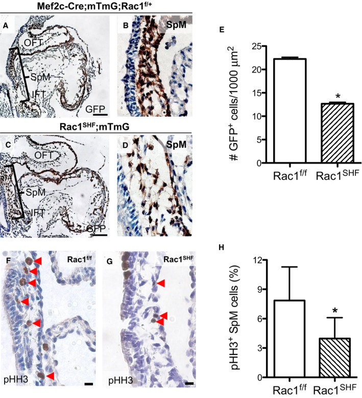
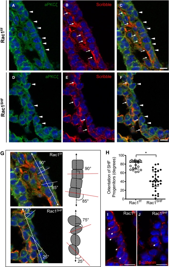
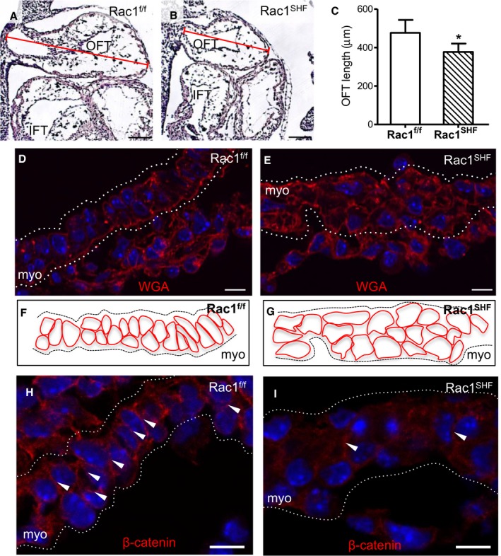
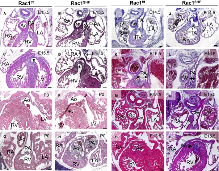
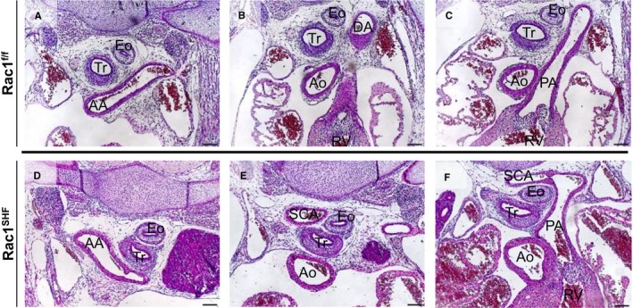
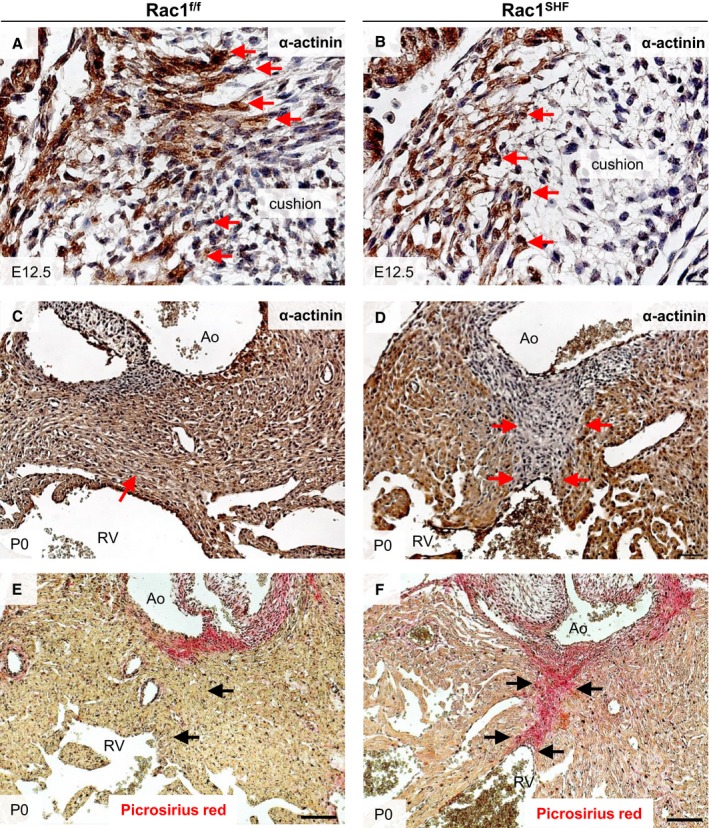
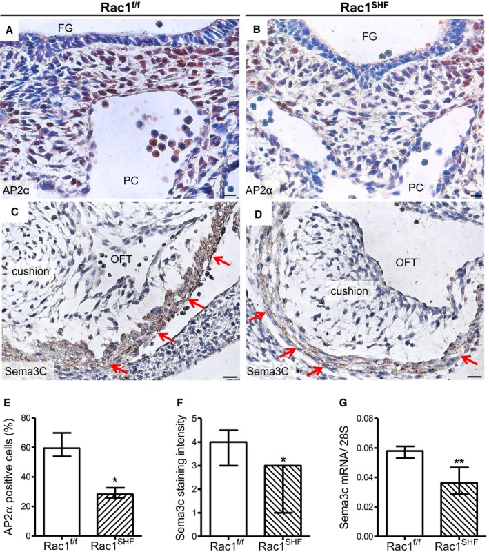
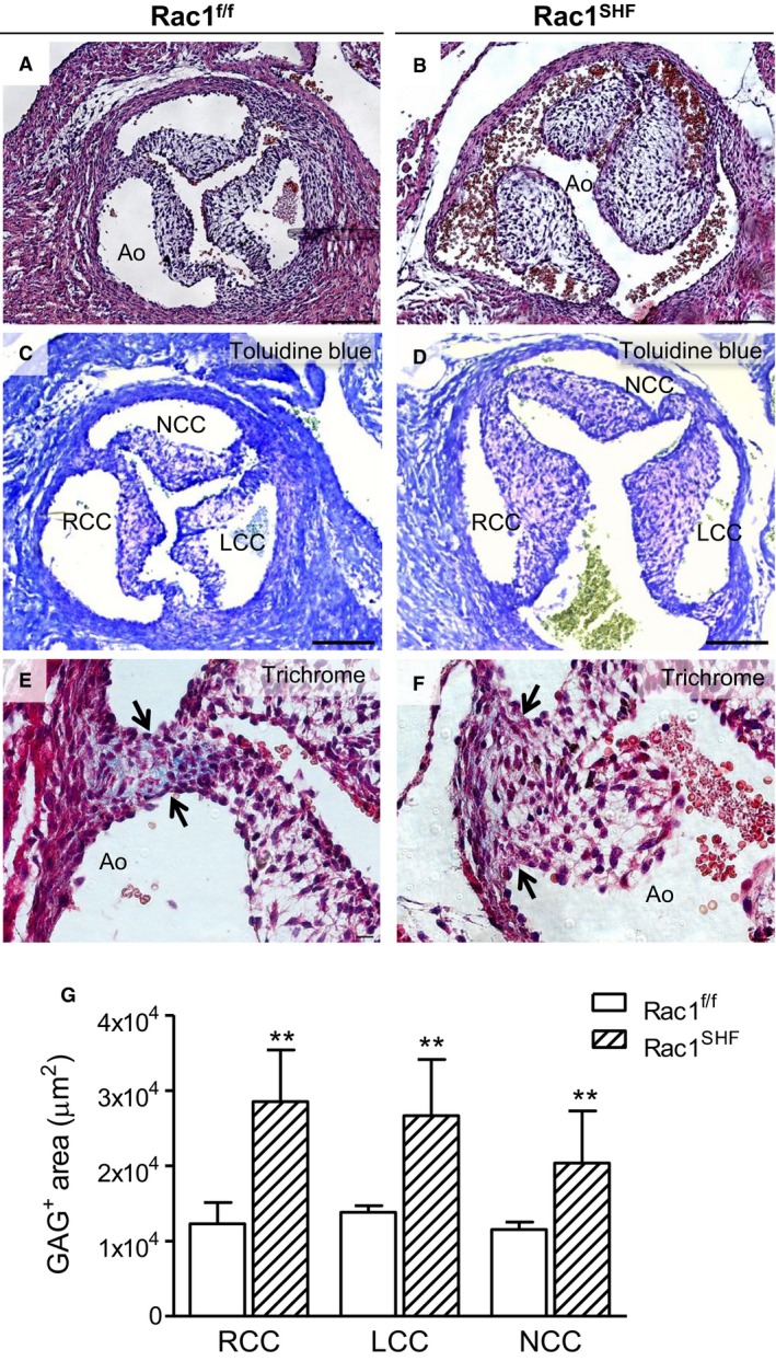
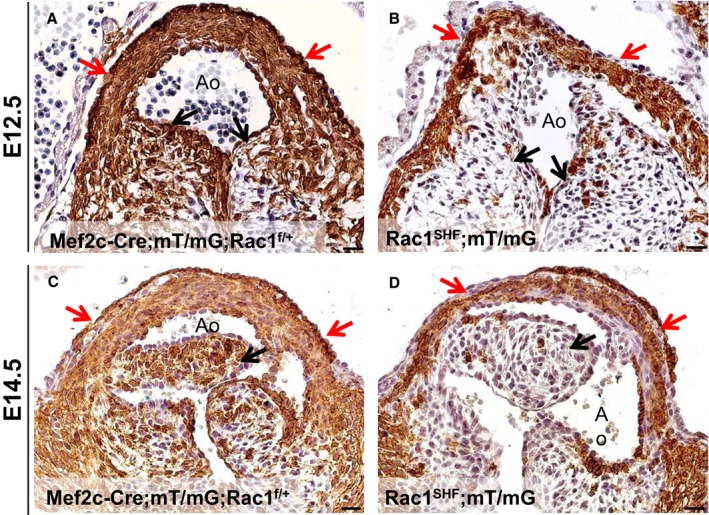
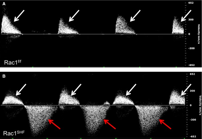
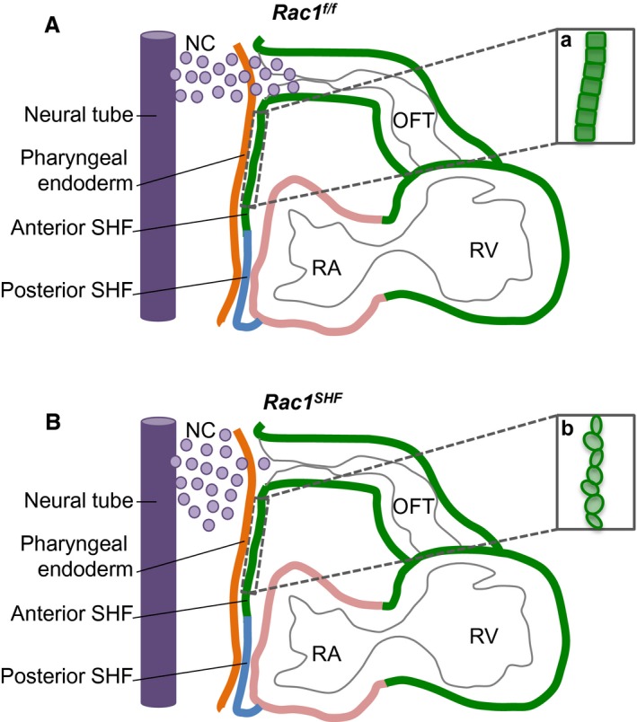
Methods and results: Using the Cre/loxP system, mice with an anterior SHF-specific deletion of Rac1 (Rac1(SHF)) were generated. Embryos were collected at various developmental time points for immunostaining and histological analysis. Intrauterine echocardiography was also performed to assess aortic valve blood flow in embryos at embryonic day 18.5. The Rac1(SHF) splanchnic mesoderm exhibited disruptions in SHF progenitor cellular organization and proliferation. Consequently, this led to a spectrum of OFT defects along with aortic valve defects in Rac1(SHF) embryos. Mechanistically, it was found that the ability of the Rac1(SHF) OFT myocardial cells to migrate into the proximal OFT cushion was severely reduced. In addition, expression of the neural crest chemoattractant semaphorin 3c was decreased. Lineage tracing showed that anterior SHF contribution to the OFT myocardium and aortic valves was deficient in Rac1(SHF) hearts. Furthermore, functional analysis with intrauterine echocardiography at embryonic day 18.5 showed aortic valve regurgitation in Rac1(SHF) hearts, which was not seen in control hearts.

Conclusions: Disruptions of Rac1 signaling in the anterior SHF results in aberrant progenitor cellular organization and defects in OFT development. Our data show Rac1 signaling to be a critical regulator of cardiac OFT formation during embryonic heart development.

Keywords: Rac1; cellular organization; congenital heart defect; outflow tract development.


Figures

Figure 1
Figure 1
Early defects in embryonic day 9.5 (E9.5) Rac1 SHF splanchnic mesoderm. Lineage tracing with Mef2c‐Cre;mT/mG showed a decreased number of GFP + SHF cells along with large acellular spaces in the splanchnic mesoderm of Rac1 SHF ;mT/mG (C and D) compared with littermate control embryos (A and B). The number of GFP + SHF progenitor cells in the splanchnic mesoderm was significantly reduced in E9.5 Rac1 SHF ;mT/mG compared with controls (E). n=4 embryos per group. Immunostaining for pHH3 in the splanchnic mesoderm (F and G) showed a reduced proliferation rate in Rac1 SHF SHF progenitors compared to Rac1 f/f controls (H). n=4 (Rac1 SHF) and n=5 (Rac1 f/f) embryos. Scale bars: 100 μm (A and C), 10 μm (B and D), 20 μm (F and G). *P<0.05 by Mann–Whitney test. GFP indicates green fluorescent protein; IFT, inflow tract; OFT, outflow tract; pHH3, phosphohistone H3; SHF, second heart field; SpM, splanchnic mesoderm.
Figure 2
Figure 2
Disrupted apicobasal cell polarity and orientation in Rac1 SHF splanchnic mesoderm. A and B, In embryonic day 9.5 (E9.5) Rac1 f/f SHF progenitors, the basolateral domain is marked by Scribble (B, arrows), and the apical domain is marked by aPKCζ (A, arrowheads). The SHF progenitors have a distinct cuboidal shape, forming an organized epithelial layer (C). Polarity is disrupted in Rac1 SHF SHF progenitors in which the Scribble‐positive basal domains (E, arrows) and aPKCζ‐positive apical domains (D, arrowheads) of individual cells are no longer aligned with neighboring cells due to the rounded morphology of the SHF progenitors (F). C, Overlay of panels A and B. F, Overlay of panels D and E. The angle of the E9.5 SHF progenitor cell long axis (dashed line) was measured relative to the axis of the dorsal pericardial wall (dashed arrow line) to obtain the degree of orientation (G). Images in panel G are from panels C and F with an overlay of schematic axis and angle measurements. H, The angle of each SHF progenitor cell in the splanchnic mesoderm was measured and plotted. *P<0.05 by Mann–Whitney test, n=4 embryos per group. Each data point represents 1 SHF progenitor cell. Active (nonphosphorylated) β‐catenin marked cell–cell junctions in E9.5 Rac1 f/f SHF progenitors (I, arrows). In comparison, cell–cell junctions were disrupted in E9.5 Rac1 SHF SHF progenitors (J). Scale bars: 10 μm (A through F, I and J). SHF indicates second heart field.
Figure 3
Figure 3
Defects in early Rac1 SHF outflow tract development. A through C, Length of the OFT at embryonic day 10.5 (E10.5) was measured in sagittal sections (A and B) and was found to be significantly shorter in Rac1 SHF compared with Rac1 f/f controls (C). * P<0.05 by Mann–Whitney test, n=4 (Rac1 SHF) and n=5 (Rac1 f/f) embryos. D and E, WGA staining to mark cell membranes of SHF cells in the OFT at E10.5 show a disorganized myocardial layer in Rac1 SHF compared with Rac1 f/f littermate controls. F and G, Schematic diagrams of panels D and E, respectively. H and I, Active (nonphosphorylated) β‐catenin staining is reduced at cell–cell junctions in E10.5 Rac1 SHF OFT myocardial cells. White arrows indicate cell–cell adhesion sites. Dotted white lines indicate boundaries of the OFT myocardial layer in panels D, E H, and I. Scale bars: 100 μm (A and B), 10 μm (D, E, H, and I). IFT indicates inflow tract; myo indicates myocardium; OFT, outflow tract; SHF, second heart field; WGA, wheat germ agglutinin.
Figure 4
Figure 4
Spectrum of OFT defects found in E14.5‐P0 Rac1 SHF hearts. Double‐outlet right ventricle was found in E15.5 Rac1 SHF hearts (A through D). The PA connects to the RV in both Rac1 SHF and Rac1 f/f hearts (A and B). The Rac1 SHF Ao (arrow) incorrectly connects to the RV compared with Rac1 f/f controls, in which the Ao connects to the LV (C and D). Overriding Ao, in which the Ao is positioned directly over a ventricular septal defect (arrow), was observed in Rac1 SHF hearts (E and F). Transposition of the great arteries was found in 1 Rac1 SHF sample in which the Ao and PA openings were switched, which resulted in the Ao connecting to the RV in Rac1 SHF and the PA connecting to the LV (G and H), respectively. PTA, in which the common OFT is not divided into aorta and pulmonary, was observed in Rac1 SHF hearts (I and J). Stenosis of the PA was observed in Rac1 SHF hearts (K and L). Aortic atresia was observed in Rac1 SHF hearts (M and N), and in 1 sample with aortic atresia, distinct aortic valves were absent (O and P). Scale bars: 100 μm. Ao indicates aorta; E, embryonic day; LA, left atrium; LV, left ventricle; OFT, outflow tract; PA, pulmonary artery; PTA, persistent truncus arteriosus; RA, right atrium; RV, right ventricle; SHF, second heart field.
Figure 5
Figure 5
Vascular rings in embryonic day 15.5 Rac1 SHF hearts. A right‐sided AA was observed in Rac1 SHF (D) hearts compared with Rac1 f/f controls (A). An aberrant retroesophageal right SCA, which arose from AA, was observed next to the Tr and Eo in Rac1 SHF hearts (E) compared with Rac1 f/f controls (B). The SCA in Rac1 SHF hearts joined with the PA, forming a vascular ring (F) in Rac1 SHF hearts compared with controls (C). Scale bars: 100 μm. AA indicates aortic arch; Ao, aorta; DA, dorsal aorta; Eo, esophagus; PA, pulmonary artery; SCA, subclavian artery; Tr, trachea.
Figure 6
Figure 6
Abnormalities in Rac1 SHF OFT myocardialization. E12.5 Rac1 SHF OFT cardiomyocytes, marked by α‐actinin immunostaining (B), exhibited a blunted morphology instead of a polarized morphology and did not extend as far into the OFT cushions compared with controls (A). Red arrows indicate invading cardiomyocytes. The proximal OFT septum remained nonmuscularized in P0 Rac1 SHF hearts (D), indicated by an absence of α‐actinin staining (arrows in D) compared with control (C), in which the septum is muscularized (arrow in C). The nonmuscularized proximal OFT septum in Rac1 SHF hearts (F) stained positive for picrosirius red (arrows in F), indicating that this tissue remained mesenchymal compared with controls (E), which had become muscularized (arrow in E). n=3 for each staining per group. Scale bars: 10 μm (A and B), 50 μm (C through F). Ao indicates aorta; E, embryonic day; OFT, outflow tract; RV, right ventricle; SHF, second heart field.
Figure 7
Figure 7
Defects in neural crest cell contribution to OFT development in Rac1 SHF hearts. A and B, AP2α immunostaining was performed to mark neural crest cells in the pharyngeal region of embryonic day 10.5 (E10.5) samples. C and D, Sema3c immunostaining (brown color) in Rac1 SHF and Rac1 f/f OFT myocardium at E11.5. E, The number of neural crest cells was significantly reduced in E10.5 Rac1 SHF pharyngeal arches compared with controls. F, Intensity of Sema3c staining in the E11.5 OFT myocardium was ranked on a scale from 1 to 5. Overall intensity of Sema3c staining was reduced in Rac1 SHF compared with Rac1 f/f controls. G, The level of Sema3c mRNA in Rac1 SHF hearts at E13.5 was significantly reduced compared with controls. * P<0.05 Mann–Whitney test. n=4 to 5 hearts (E and F) and n=7 to 8 hearts (G) per group. Scale bars: 20 μm (A and B), 10 μm (C and D). FG indicates foregut; OFT, outflow tract; PC, pericardial cavity; Sema3c, semaphorin 3c; SHF, second heart field.
Figure 8
Figure 8
Aortic valve defects in P0 Rac1 SHF hearts. More than 30% (9 of 28) of P0 Rac1 SHF hearts exhibited thick aortic valve leaflets (B) compared with the thin, remodeled valves of controls (A). Toluidine blue staining showed that GAG (light purple color) occupies the acellular space of Rac1 SHF and littermate valve leaflets (C and D). Masson's trichrome staining in Rac1 f/f aortic valves showed collagen in the commissure of valve leaflets (E, arrows), which was absent in Rac1 SHF aortic valves (F, arrows). The GAG‐positive area (light purple color) in each valve leaflet (C and D) was quantified in (G). **P<0.01 by Mann–Whitney test, n=5 to 6 hearts per group. Scale bars: 100 μm (A through D), 10 μm (E and F). Ao indicates aorta; GAG, glycosaminoglycans; LCC, left coronary cusp; NCC, noncoronary cusp; RCC, right coronary cusp; SHF, second heart field.
Figure 9
Figure 9
Decreased SHF contribution to Rac1 SHF OFT. Fate mapping with mT/mG reporter shows a decreased SHF progenitor contribution to the OFT myocardium (red arrows in B and D) in E12.5 and E14.5 Rac1 SHF hearts compared with controls (red arrows in A and C). SHF contribution to the valve leaflets was also severely reduced in Rac1 SHF hearts compared with controls (black arrows, A through D). Paraffin sections were immunostained with anti‐GFP. Scale bars: 20 μm. n=3 for each time point per group. Ao indicates aorta; E, embryonic day; OFT, outflow tract; SHF, second heart field.
Figure 10
Figure 10
Aortic valve regurgitation in Rac1 SHF hearts. Intrauterine echocardiography at embryonic day 18.5 showed that Rac1 SHF hearts had severe aortic regurgitation during diastole (B, red arrows), which is not observed in Rac1 f/f littermates (A). White arrows in (A and B) indicate forward blood flow during systole. Shown are representatives of 5 fetuses per group. SHF indicates second heart field.
Figure 11
Figure 11
Schematic diagram of midsagittal section of embryonic day 9.5 (E9.5) splanchnic mesoderm. At E9.5, the Rac1 f/f anterior SHF progenitors in the splanchnic mesoderm are organized into a polarized epithelium (a). Chemotactic signals, such as semaphorin 3c, secreted by the OFT myocardium act as axonal guidance cues for migration of cardiac NC cells from the neural tube (A). In Rac1 SHFembryos, the anterior SHF progenitors are rounded and disorganized, displaying a loss of polarized epithelium characteristics (b). Expression of chemotactic signals is reduced, resulting in decreased migration of neural cells into the OFT (B). NC indicates neural crest; OFT, outflow tract; RA, right atrium; RV, right ventricle; SHF, second heart field.

References

    1. Bruneau BG. The developmental genetics of congenital heart disease. Nature. 2008;451:943–948. - PubMed
    1. Jenkins KJ, Gauvreau K, Newburger JW, Spray TL, Moller JH, Iezzoni LI. Consensus‐based method for risk adjustment for surgery for congenital heart disease. J Thorac Cardiovasc Surg. 2002;123:110–118. - PubMed
    1. Bryant DM, Mostov KE. From cells to organs: building polarized tissue. Nat Rev Mol Cell Biol. 2008;9:887–901. - PMC - PubMed
    1. Phillips HM, Rhee HJ, Murdoch JN, Hildreth V, Peat JD, Anderson RH, Copp AJ, Chaudhry B, Henderson DJ. Disruption of planar cell polarity signaling results in congenital heart defects and cardiomyopathy attributable to early cardiomyocyte disorganization. Circ Res. 2007;101:137–145. - PubMed
    1. Henderson DJ, Phillips HM, Chaudhry B. Vang‐like 2 and noncanonical Wnt signaling in outflow tract development. Trends Cardiovasc Med. 2006;16:38–45. - PubMed

Publication types

MeSH terms