

Endostar enhances the antitumor effects of radiation by affecting energy metabolism and alleviating the tumor microenvironment in a Lewis lung carcinoma mouse model
- PMID: 26722291
- PMCID: PMC4665656
- DOI: 10.3892/ol.2015.3679
Endostar enhances the antitumor effects of radiation by affecting energy metabolism and alleviating the tumor microenvironment in a Lewis lung carcinoma mouse model
Abstract
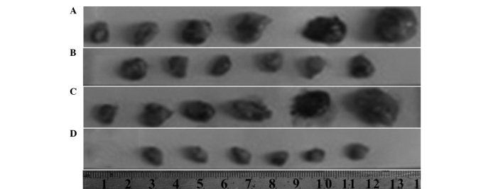
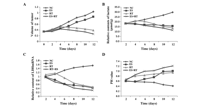
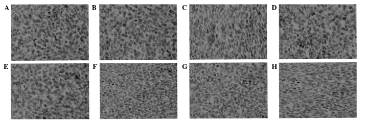
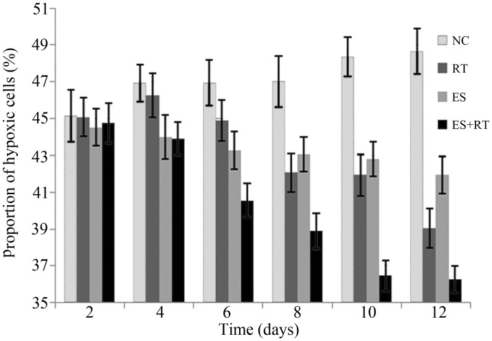
Lung cancer is a leading cause of morbidity and mortality. Previous studies have identified that an improvement in treatment efficacy was achieved using Endostar; however, the role of Endostar in lung cancer remains poorly understood. The present study investigated whether the enhanced antitumor effects of Endostar in combination with radiation involved changes in the metabolism and microenvironment in non-small cell lung cancer. A Lewis lung carcinoma mouse model was used, including the control, Endostar (ES), radiotherapy (RT) and Endostar plus radiotherapy (ES + RT) groups. The tumor inhibition rates and growth were described based on changes in tumor volume. In addition, ultraviolet enzymatic analysis was performed to determine the lactate level and reverse transcription-polymerase chain reaction was used to measure the mRNA expression of lactate dehydrogenase (LDH). A Meph-3 pH meter was used to detect the ranges of tumor interstitial tissue pH, and immunohistochemical analysis was adopted to examine hypoxia within the tumor microenvironment. The tumor inhibition rate of the ES + RT group was significantly higher compared with the other three groups (P<0.05). Following treatment, the lactate levels decreased in all three treatment groups compared with the control, particularly in the ES + RT group (P<0.05). Reduced LDH expression and hypoxic fraction in the tumor microenvironment were also observed in the ES + RT group (P<0.05). Furthermore, changes from acidic to alkaline pH in the tumor microenvironment were detected in the ES + RT group. The present study suggested that Endostar is involved in the regulation of metabolism and tumor microenvironment hypoxia, which may be responsible for the enhanced antitumor effect of Endostar in combination with radiotherapy.
Keywords: endostar; hypoxia; lactate; lung cancer; radiotherapy.
Figures
Similar articles
-
The effect of combining Endostar with radiotherapy on blood vessels, tumor-associated macrophages, and T cells in brain metastases of Lewis lung cancer.Transl Lung Cancer Res. 2020 Jun;9(3):745-760. doi: 10.21037/tlcr-20-500. Transl Lung Cancer Res. 2020. PMID: 32676336 Free PMC article.
-
PDGFR-β inhibitor slows tumor growth but increases metastasis in combined radiotherapy and Endostar therapy.Biomed Pharmacother. 2018 Mar;99:615-621. doi: 10.1016/j.biopha.2018.01.095. Epub 2018 Feb 20. Biomed Pharmacother. 2018. PMID: 29653486
-
Antitumor activity of Endostar combined with radiation against human nasopharyngeal carcinoma in mouse xenograft models.Oncol Lett. 2012 Nov;4(5):976-980. doi: 10.3892/ol.2012.856. Epub 2012 Aug 8. Oncol Lett. 2012. PMID: 23162635 Free PMC article.
-
Antitumor effects of Endostar(rh-endostatin) combined with gemcitabine in different administration sequences to treat Lewis lung carcinoma.Cancer Manag Res. 2019 Apr 23;11:3469-3479. doi: 10.2147/CMAR.S192868. eCollection 2019. Cancer Manag Res. 2019. PMID: 31114380 Free PMC article.
-
Emerging Bismuth Chalcogenides Based Nanodrugs for Cancer Radiotherapy.Front Pharmacol. 2022 Feb 18;13:844037. doi: 10.3389/fphar.2022.844037. eCollection 2022. Front Pharmacol. 2022. PMID: 35250594 Free PMC article. Review.
Cited by
-
The Preventive Effect of Endostar on Radiation-induced Pulmonary Fibrosis.Curr Mol Med. 2024;24(5):610-619. doi: 10.2174/1566524023666230406134640. Curr Mol Med. 2024. PMID: 37038709
-
Endostar acts as a pneumonitis protectant in patients with locally advanced non-small cell lung cancer receiving concurrent chemoradiotherapy.BMC Cancer. 2024 Feb 23;24(1):257. doi: 10.1186/s12885-024-12001-6. BMC Cancer. 2024. PMID: 38395838 Free PMC article.
-
Anti-tumor effect of local injectable hydrogel-loaded endostatin alone and in combination with radiotherapy for lung cancer.Drug Deliv. 2021 Dec;28(1):183-194. doi: 10.1080/10717544.2020.1869864. Drug Deliv. 2021. PMID: 33427520 Free PMC article.
-
Heterogeneity of circulating tumor cell dissemination and lung metastases in a subcutaneous Lewis lung carcinoma model.Biomed Opt Express. 2020 Jun 8;11(7):3633-3647. doi: 10.1364/BOE.395289. eCollection 2020 Jul 1. Biomed Opt Express. 2020. PMID: 33014556 Free PMC article.
-
Metabolic Interplay between Tumour Cells and Cancer-Associated Fibroblasts (CAFs) under Hypoxia versus Normoxia.Malays J Med Sci. 2018 May;25(3):7-16. doi: 10.21315/mjms2018.25.3.2. Epub 2018 Jun 28. Malays J Med Sci. 2018. PMID: 30899183 Free PMC article. Review.
References
-
- No authors listed: Cancer facts and figures 2004. American Cancer Society; Atlanta, GA: 2004.
LinkOut - more resources
Full Text Sources
Other Literature Sources