

Increasing the Inflammatory Competence of Macrophages with IL-6 or with Combination of IL-4 and LPS Restrains the Invasiveness of Pancreatic Cancer Cells
- PMID: 26722359
- PMCID: PMC4679380
- DOI: 10.7150/jca.12923
Increasing the Inflammatory Competence of Macrophages with IL-6 or with Combination of IL-4 and LPS Restrains the Invasiveness of Pancreatic Cancer Cells
Abstract
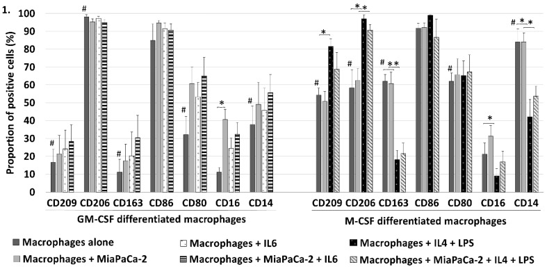
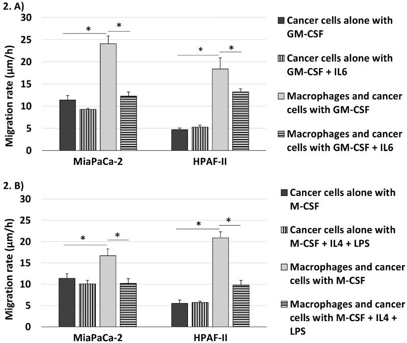
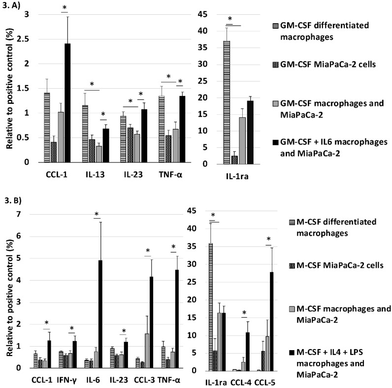
Recent studies suggest that pro-inflammatory type M1 macrophages inhibit tumor progression and that anti-inflammatory M2 macrophages enhance it. The aim of this study was to examine the interaction of type M1 and M2 macrophages with pancreatic cancer cells. We studied the migration rate of fluorescein stained pancreatic cancer cells on Matrigel cultured alone or with Granulocyte-Macrophage Colony Stimulating Factor (GM-CSF) differentiated macrophages or with Macrophage Colony Stimulating Factor (M-CSF) differentiated macrophages, skewing the phenotype towards pro- and anti-inflammatory direction, respectively. Macrophage differentiation was assessed with flow cytometry and the cytokine secretion in cell cultures with cytokine array. Both GM-CSF and M-CSF differentiated macrophages increased the migration rate of primary pancreatic adenocarcinoma cell line (MiaPaCa-2) and metastatic cell line (HPAF-II). Stimulation with IL6 or IL4+LPS reversed the macrophages' increasing effect on the migration rate of MiaPaCa-2 completely and partly of HPAF-II. Co-culture with MiaPaCa-2 reduced the inflammatory cytokine secretion of GM-CSF differentiated macrophages. Co-culture of macrophages with pancreatic cancer cells seem to change the inflammatory cytokine profile of GM-CSF differentiated macrophages and this might explain why also GM-CSF differentiated macrophages promoted the invasion. Adding IL6 or IL4+LPS to the cell culture with MiaPaCa-2 and GM-CSF or M-CSF differentiated macrophages increased the secretion of inflammatory cytokines and this could contribute to the reversion of the macrophage induced increase of cancer cell migration rate.
Keywords: M1; M2; invasion; macrophages; pancreatic adenocarcinoma.
Conflict of interest statement
Competing Interests: The authors have declared that no competing interest exists.
Figures
References
-
- National Cancer Institute, SEER Cancer Statistics Review, 1975-2012. http://seer.cancer.gov/csr/1975_2012/browse_csr.php?sectionSEL=22&pageSE....
-
- Ekbom A, McLaughlin JK, Nyren O. Pancreatitis and the risk of pancreatic cancer. N Engl J Med. 1993;329:1502–1503. - PubMed
-
- Zhou D, Huang C, Lin Z, Zhan S, Kong L, Fang C. et al. Macrophage polarization and function with emphasis on the evolving roles of coordinated regulation of cellular signaling pathways. Cell Signal. 2014;26:192–197. doi: 10.1016/j.cellsig.2013.11.004. - PubMed
LinkOut - more resources
Full Text Sources
Other Literature Sources
Research Materials
