. 2016 Mar 1:496:43-9.
doi: 10.1016/j.ab.2015.11.027. Epub 2015 Dec 23.

A continuous spectrophotometric enzyme-coupled assay for deoxynucleoside triphosphate triphosphohydrolases

Affiliations

A continuous spectrophotometric enzyme-coupled assay for deoxynucleoside triphosphate triphosphohydrolases

Deepa Singh et al. Anal Biochem. .

Abstract

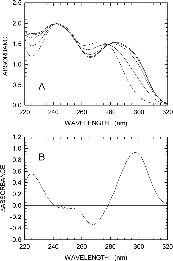
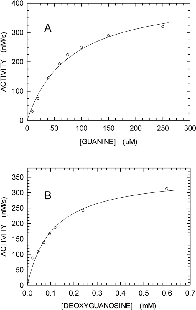
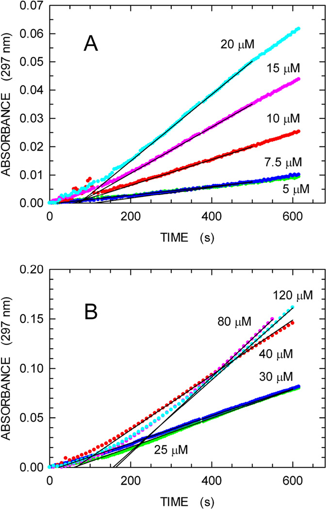
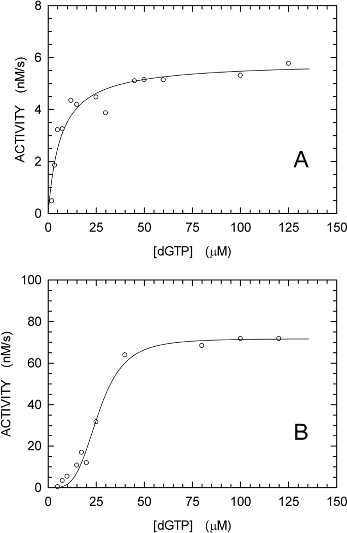
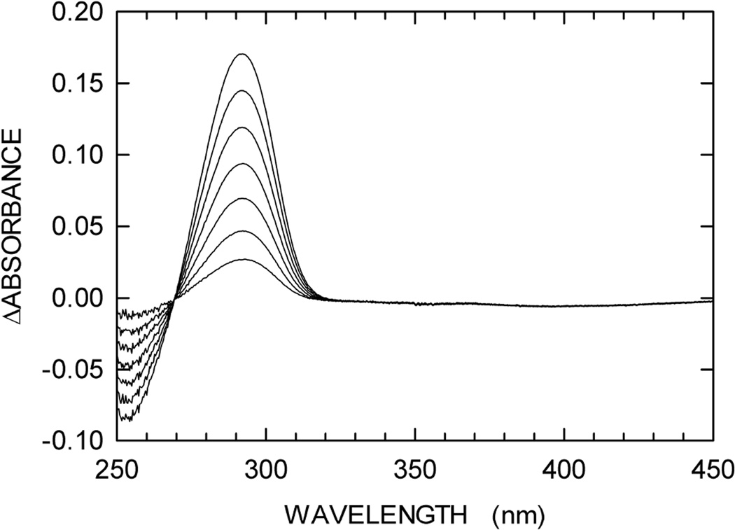
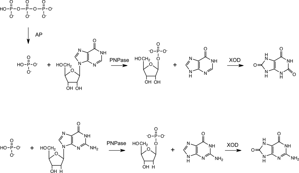
We describe a continuous, spectrophotometric, enzyme-coupled assay useful to monitor reactions catalyzed by nucleoside triphosphohydrolases. In particular, using Escherichia coli deoxynucleoside triphosphohydrolase (Dgt), which hydrolyzes dGTP to deoxyguanosine and tripolyphosphate (PPPi) as the enzyme to be tested, we devised a procedure relying on purine nucleoside phosphorylase (PNPase) and xanthine oxidase (XOD) as the auxiliary enzymes. The deoxyguanosine released by Dgt can indeed be conveniently subjected to phosphorolysis by PNPase, yielding deoxyribose-1-phosphate and guanine, which in turn can be oxidized to 8-oxoguanine by XOD. By this means, it was possible to continuously detect Dgt activity at 297 nm, at which wavelength the difference between the molar extinction coefficients of 8-oxoguanine (8000 M(-1) cm(-1)) and guanine (1090 M(-1) cm(-1)) is maximal. The initial velocities of Dgt-catalyzed reactions were then determined in parallel with the enzyme-coupled assay and with a discontinuous high-performance liquid chromatography (HPLC) method able to selectively detect deoxyguanosine. Under appropriate conditions of excess auxiliary enzymes, the activities determined with our continuous enzyme-coupled assay were quantitatively comparable to those observed with the HPLC method. Moreover, the enzyme-coupled assay proved to be more sensitive than the chromatographic procedure, permitting reliable detection of Dgt activity at low dGTP substrate concentrations.

Keywords: 8-Oxoguanine; Deoxynucleoside triphosphatase; Enzyme-coupled assay; Escherichia coli Dgt; Purine nucleoside phosphorylase; Xanthine oxidase.


Figures

Fig. 1
Fig. 1
Oxidation of guanine by xanthine oxidase (XOD). The reactions were performed as described in Materials and methods. (A) The absorption spectrum of 0.3 mM guanine was first determined in 100 mM Tris-HCl pH 8 (black line). XOD was then added to the cuvette, and spectra were recorded at 5, 10, 20, and 40 min after enzyme addition (green, blue, red, and dark-green lines, respectively). A reference cuvette was used containing 100 mM Tris-HCl pH 8 and XOD. (B) Difference between the spectra recorded before and after 40 min of XOD addition.
Fig. 2
Fig. 2
Initial velocities of reactions catalyzed by XOD and PNPase. (A) Oxidation of guanine by XOD as a function of substrate concentration. The continuous line represents the best fit to the Michaelis-Menten equation. (B) Phosphorolysis of deoxyguanosine by PNPase as a function of substrate concentration. The continuous line represents the best fit to the Michaelis-Menten equation.
Fig. 3
Fig. 3
Enzyme-coupled assay for formation of dG from dGTP. The reaction contained dGTP substrate, Dgt enzyme, Purine Nucleoside Phosphorylase and Xanthine Oxidase, as described in Materials and Methods. The reaction was monitored by increase in absorbance at 297 nm. The concentration of dGTP was varied from 2 to 120 µM. Panels A and B show the reactions for the 2–20 µM and 25–120 µM range, respectively. For each reaction, the zero-order reaction velocity (continuous black lines) was determined and used to derive the kinetics parameters of Fig. 4B. The lag-time before steady-state intermediate concentrations are reached, as observed here, is well known for enzyme-coupled assays [29].
Fig. 4
Fig. 4
dGTPase activity of E. coli Dgt measured using two different techniques. (A) HPLC method. The Dgt (hexamer) concentration was 0.2 nM; the kcat was calculated at 29.2 ± 1.1 s−1. (B) Enzyme-coupled assay. The protein concentration used was 4 nM; the kcat was calculated at 17.9 ± 0.6 s−1. The data were fitted to the Hill equation [23]. For the curve of Fig. 4B, an N value > 1 was obtained, indicating a cooperative mechanism for dGTP hydrolysis.
Fig. 5
Fig. 5
Glycosyltransferase activity of PNPase. An assay mixture containing 100 mM Tris (pH 8), 0.15 mM thymine, 0.25 mM inosine, and 500 mU/mL of PNPase was prepared, added to sample and reference cuvettes to record a baseline in the 250–450 nm wavelength interval. 500 mU/mL of XOD were then added to the sample cuvette, and difference spectra were recorded every 30 min after enzyme addition.
Scheme 1
Scheme 1
Reaction paths starting from the products (PPPi and dG) released by the catalytic action of Dgt yielding spectrophotometrically detectable compounds. Upper path, the tripolyphosphate (PPPi) produced by Dgt is hydrolyzed by alkaline phosphatase (AP), yielding orthophosphate (Pi), which, in the presence of inosine and PNPase, is converted to ribose-1-phosphate and hypoxanthine. Finally, hypoxanthine is oxidized by XOD, yielding uric acid. Lower path, the deoxyguanosine released by Dgt is phosphorolysed by PNPase, producing deoxyribose-1-phosphate and guanine. Guanine is then converted by XOD into 8-oxoguanine.

References

    1. Brignole EJ, Ando N, Zimanyi CM, Drennan CL. The prototypic class Ia ribonucleotide reductase from Escherichia coli : still surprising after all these years. Biochem. Soc. Trans. 2012;40:523–530. - PMC - PubMed
    1. Ahmad MF, Dealwis CG. The structural basis for the allosteric regulation of ribonucleotide reductase. Prog. Mol. Biol. Transl. Sci. 2013;177:389–410. - PMC - PubMed
    1. Lascu I, Gonin P. The catalytic mechanism of nucleoside diphosphate kinases. J. Bioenerg. Biomembr. 2000;32:237–246. - PubMed
    1. Lascu L, Giartosio A, Ransac S, Erent M. Quaternary structure of nucleoside diphosphate kinases. J. Bioenerg. Biomembr. 2000;32:227–236. - PubMed
    1. Bernard MA, Ray NB, Olcott MC, Hendricks SP, Mathews CK. Metabolic functions of microbial nucleoside diphosphate kinases. J. Bioenerg. Biomembr. 2000;32:259–267. - PubMed

Publication types

MeSH terms

LinkOut - more resources