. 2016 Jan 4:6:18507.
doi: 10.1038/srep18507.

Faecalibacterium prausnitzii A2-165 has a high capacity to induce IL-10 in human and murine dendritic cells and modulates T cell responses

Affiliations

Faecalibacterium prausnitzii A2-165 has a high capacity to induce IL-10 in human and murine dendritic cells and modulates T cell responses

Oriana Rossi et al. Sci Rep. .

Abstract

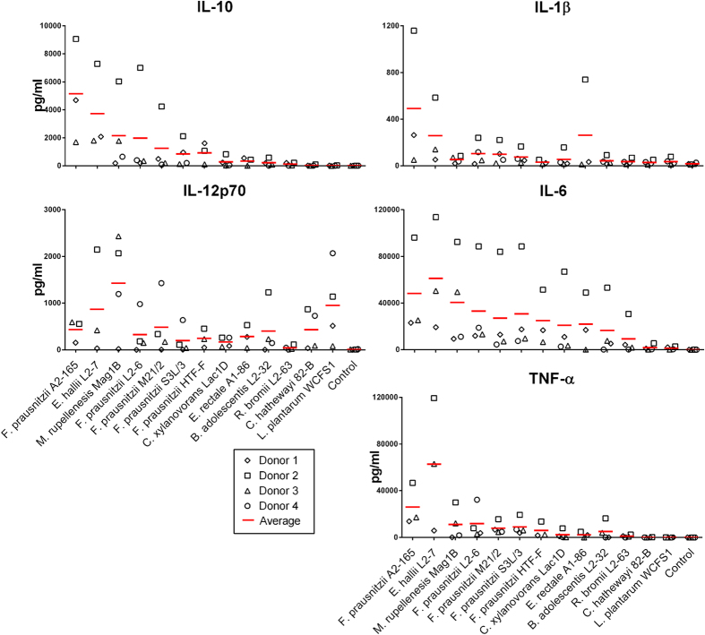
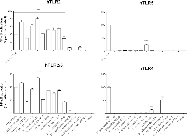
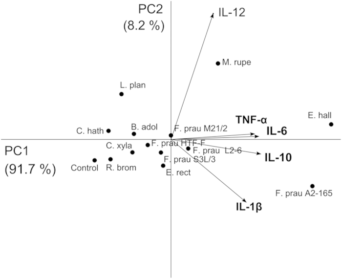
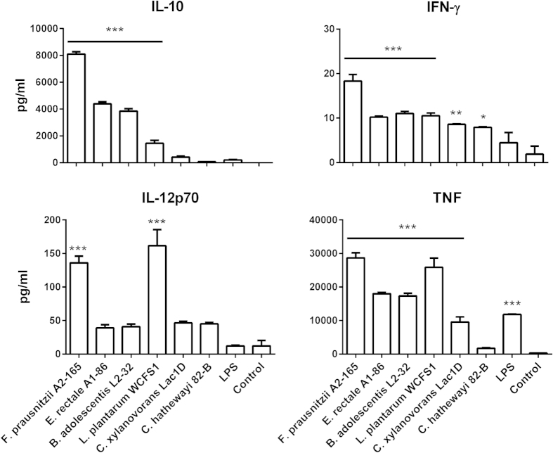
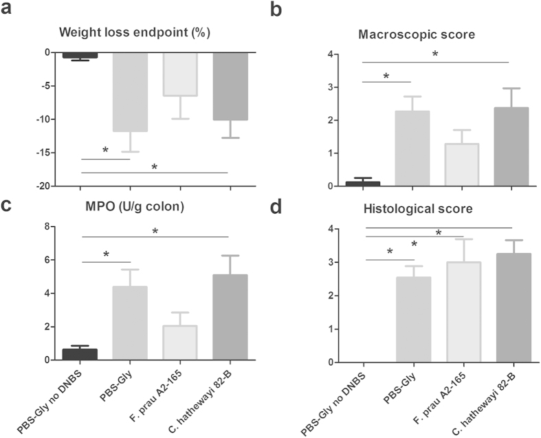
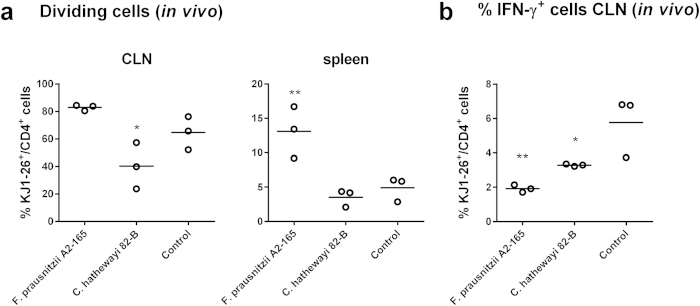
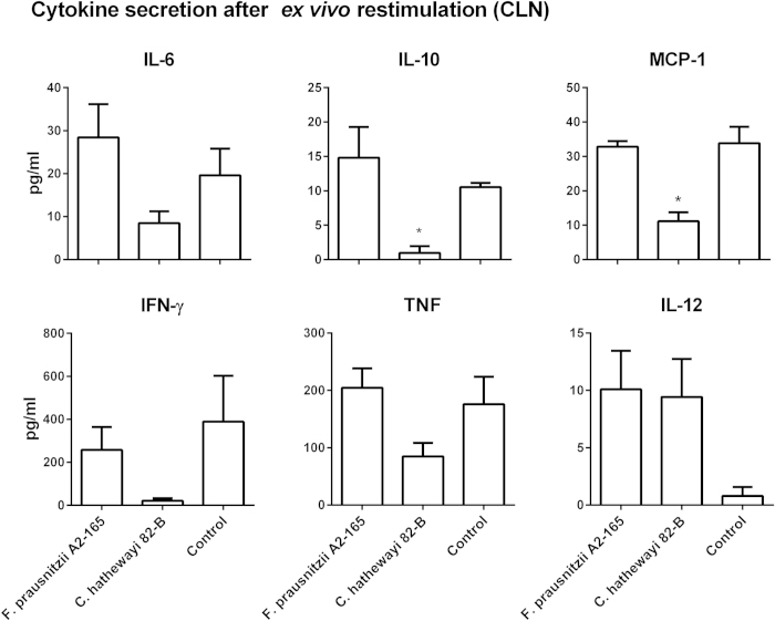
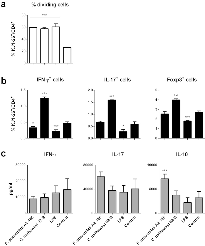
Faecalibacterium prausnitzii strain A2-165 was previously reported to have anti-inflammatory properties and prevent colitis in a TNBS model. We compared the immunomodulatory properties of strain A2-165 to four different F. prausnitzii isolates and eight abundant intestinal commensals using human dendritic cells (DCs) and mouse BMDCs in vitro. Principal component analysis revealed that the cytokine response to F. prausnitzii A2-165 is distinct from the other strains in eliciting high amounts of IL-10 secretion. The mouse DNBS model of relapsing IBD was used to compare the protective effects of F. prausnitzii A2-165 and Clostridium hathewayi, a low secretor of IL-10, on the Th1-driven inflammatory response to DNBS; attenuation of disease parameters was only observed with F. prausnitzii. In an in vivo mouse model of nasal tolerance to ovalbumin, F. prausnitzii A2-165 enhanced ovalbumin-specific T cell proliferation and reduced the proportion of IFN-γ(+) T cells in CLNs. Similarly, in vitro F. prausnitzii A2-165 stimulated BMDCs increased ovalbumin-specific T cell proliferation and reduced the number of IFN-γ(+) T cells. These mechanisms may contribute to the anti-inflammatory effects of F. prausnitzii in colitis and support the notion that this abundant bacterium might contribute to immune homeostasis in the intestine via its anti-inflammatory properties.


Figures

Figure 1
Figure 1. Cytokine secretion by hDCs. IL-10, IL-12p70, TNF-α, IL-1β and IL-6 were measured in the supernatant of hDCs from 3 to 4 donors after 48 h of incubation with the bacteria (bacterium to DC ratio 10:1, 106 DCs/well in 24w plate).
Each donor is represented with a different symbol and the red lines indicate the average of the donors.
Figure 2
Figure 2. TLR signalling properties of the commensal bacteria. NF-κB activation was measured using a luminescence reporter in HEK293 cell lines expressing TLR2, TLR2/6, TLR5 and TLR4 after incubation with the bacteria (bacterium: cell ratio, 10:1).
NF-κB activation is expressed as percentage of the positive control. Error bars represent SEM, n = 6, *indicates p < 0.05, **p < 0.01, ***p < 0.001 compared to the control.
Figure 3
Figure 3. Principal component analysis (PCA) of the cytokine profiles induced by the commensal bacteria in hDCs.
Each dot represents the average of cytokine levels of a commensal strain in hDCs. The variables (level of cytokines) are represented by vectors. The direction and length of a vector indicates how the variable contributes to the two principal components in the plot. (F. prau, F. prausnitzii; R. brom, R. bromii L2-63; E. rect, E. rectale A1-86; E. hall, E. halli L2-7; M. rupe, M. rupellensis Mag1B; C. hath, C. hathewayi 82-B; C. xyla, C. xylanovorans Lac1D; L. plan, L. plantarum WCFS1).
Figure 4
Figure 4. Cytokine secretion by mouse BMDCs. IL-10, IL-12p70, TNF and IFN-γ were measured in the supernatant of BMDCs (5 × 105 BMDCs/well in 24w plate) after 24 h of incubation with bacteria (bacterium: BMDC, 10:1).
Error bars represent SEM, n = 3, ***indicates p < 0.001, **p < 0.01, *p < 0.05 compared to the control.
Figure 5
Figure 5. Effects of F. prausnitzii A2-165 and C. hathewayi 82-B on DNBS induced colitis.
Percentage of weight loss at the end of the experiment (a), macroscopic scores (b), MPO activity (c) and histological scores (d) in control non-inflamed (PBS-Gly no DNBS), control inflamed (PBS-Gly), F. prausnitzii A2-165, C. hathewayi 82-B treated mice. Error bars represent SEM, n = 8, *indicates p < 0.05 compared to the control PBS-DNBS.
Figure 6
Figure 6. Percentage of dividing and IFN-γ+ OVA-T cells in vivo.
CFSE labelled naive OVA-T cells (KJ1-26+/CD4+) were adoptively transferred in BALB/c mice, after 24 h, mice were administered i.n. with bacteria plus OVA and after additional 72 h, OVA-T cells were isolated from cervical lymph nodes (CLNs) and spleens and analysed. (a) Percentage of dividing OVA-T cells (KJ1-26+/CD4+) isolated from CLNs or spleens. (b) Percentage of IFN-γ+ OVA-T cells (KJ1-26+/CD4+) isolated from CLNs. *indicates p < 0.05, **indicates p < 0.01 compared to the control administered OVA alone.
Figure 7
Figure 7. Cytokine secretion by OVA-T cells after ex vivo re-stimulation.
CFSE labelled naive OVA-T cells were adoptively transferred in BALB/c mice, after 24 h, mice were administered i.n. with bacteria plus OVA and after additional 72 h, OVA-T cells were isolated from CLNs and re-stimulated with OVA for additional 24 h and then analysed. *indicates p < 0.05 compared to the control administered OVA alone.
Figure 8
Figure 8. OVA-T cells (KJ1-26+/CD4+) division (a), IFN-γ+, IL-17+, Foxp3+ cells (b) and cytokine secretion (c) after incubation with BMDCs pre-stimulated with bacteria (bacterium: BMDC, 10: 1).
BMDCs (5 × 104 BMDCs/well in 96w plate) were loaded with OVA (0.5 mg/ml) and cultured in the presence or absence of the bacteria or LPS and after 24 h, CFSE labelled OVA-T cells (5 × 105 T cells/well) were added, after 72 h cytokine secretion in the supernatant and intracellular markers were measured. Error bars represent SEM, n = 3, *** indicates p < 0.001 compared to the control, *p < 0.05 compared to the control. Intracellular IL-10 staining was negative for all samples.

References

    1. Whitman W. B., Coleman D. C. & Wiebe W. J. Prokaryotes: the unseen majority. Proc Natl Acad Sci USA 95, 6578–6583 (1998). - PMC - PubMed
    1. Lopez-Siles M. et al. Cultured representatives of two major phylogroups of human colonic Faecalibacterium prausnitzii can utilize pectin, uronic acids, and host-derived substrates for growth. Applied and environmental microbiology 78, 420–428, doi: 10.1128/AEM.06858-11 (2012). - DOI - PMC - PubMed
    1. Sekirov I., Russell S. L., Antunes L. C. & Finlay B. B. Gut microbiota in health and disease. Physiological reviews 90, 859–904, doi: 10.1152/physrev.00045.2009 (2010). - DOI - PubMed
    1. Kiyono H., McGhee J. R., Wannemuehler M. J. & Michalek S. M. Lack of oral tolerance in C3H/HeJ mice. J Exp Med 155, 605–610 (1982). - PMC - PubMed
    1. Tanaka K. & Ishikawa H. Role of intestinal bacterial flora in oral tolerance induction. Histology and histopathology 19, 907–914 (2004). - PubMed

Publication types

MeSH terms

LinkOut - more resources