. 2016 Jan 19;32(2):551-9.
doi: 10.1021/acs.langmuir.5b03445. Epub 2016 Jan 4.

Nonfouling NTA-PEG-Based TEM Grid Coatings for Selective Capture of Histidine-Tagged Protein Targets from Cell Lysates

Affiliations

Nonfouling NTA-PEG-Based TEM Grid Coatings for Selective Capture of Histidine-Tagged Protein Targets from Cell Lysates

Christopher J Benjamin et al. Langmuir. .

Abstract

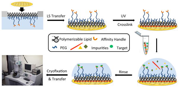
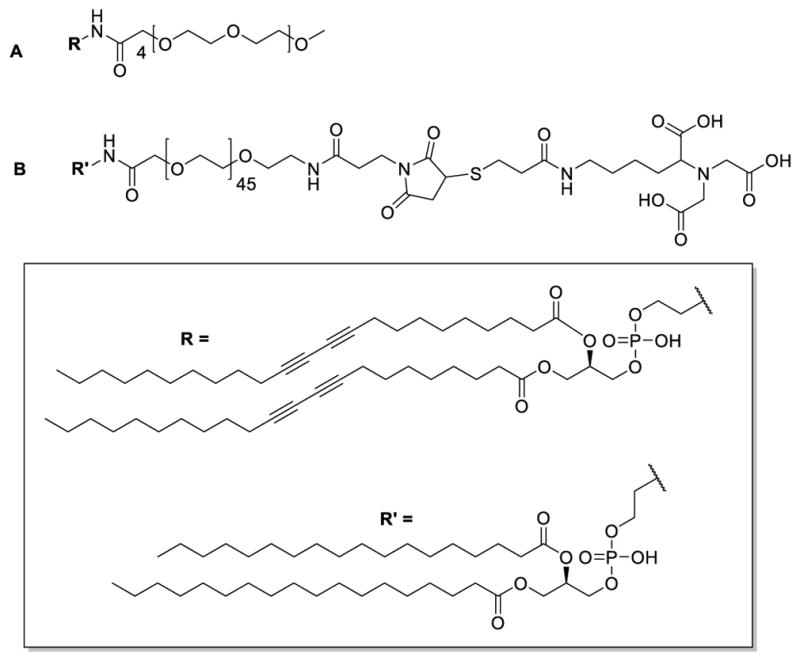
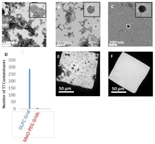
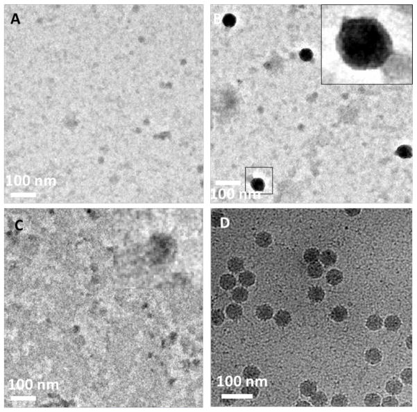
We report the preparation and performance of TEM grids bearing stabilized nonfouling lipid monolayer coatings. These films contain NTA capture ligands of controllable areal density at the distal end of a flexible poly(ethylene glycol) 2000 (PEG2000) spacer to avoid preferred orientation of surface-bound histidine-tagged (His-tag) protein targets. Langmuir-Schaefer deposition at 30 mN/m of mixed monolayers containing two novel synthetic lipids-1,2-distearoyl-sn-glycero-3-phosphoethanolamine-N-[(5-amido-1-carboxypentyl)iminodiacetic acid]polyethylene glycolamide 2000) (NTA-PEG2000-DSPE) and 1,2-(tricosa-10',12'-diynoyl)-sn-glycero-3-phosphoethanolamine-N-(methoxypolyethylene glycolamide 350) (mPEG350-DTPE)-in 1:99 and 5:95 molar ratios prior to treatment with a 5 min, 254 nm light exposure was used for grid fabrication. These conditions were designed to limit nonspecific protein adsorption onto the stabilized lipid coating by favoring the formation of a mPEG350 brush layer below a flexible, mushroom conformation of NTA-PEG2000 at low surface density to enable specific immobilization and random orientation of the protein target on the EM grid. These grids were then used to capture His6-T7 bacteriophage and RplL from cell lysates, as well as purified His8-green fluorescent protein (GFP) and nanodisc solubilized maltose transporter, His6-MalFGK2. Our findings indicate that TEM grid supported, polymerized NTA lipid monolayers are capable of capturing His-tag protein targets in a manner that controls their areal densities, while efficiently blocking nonspecific adsorption and limiting film degradation, even upon prolonged detergent exposure.


Figures

Figure 1
Figure 1
Conceptual diagram showing stabilized monolayer affinity grid fabrication and utilization. (Top left) A fluid-phase mixed lipid monolayer comprised of Ni2+:NTA-PEG2000-DSPE and mPEG350-DTPE (1:99 or 5:95 mol:mol) was compressed until the mPEG350-DTPE component entered the brush regime and the diyne lipid chains were fully condensed; (Top center) the condensed lipid monolayer film was deposited onto carbon-coated TEM grids via Langmuir-Schaefer (LS) transfer; (Top right) photopolymerization of the LS film, followed by Ni2+ activation, produces a grid with a stable affinity capture coating; (Bottom right) solutions containing the His-tag protein of interest are deposited onto the coated grids, either as a clarified cell lysate or as a purified protein sample; (Bottom center) blotting and rinsing of the sample removes non-target material prior to (Bottom left) cryofixation (or negative staining) and sample imaging.
Figure 2
Figure 2
Structures of (A) mPEG350-DTPE and (B) NTA-PEG2000-DSPE.
Figure 3
Figure 3
Comparative performance of uncoated (bare carbon, glow-discharge), DLPC, and mPEG350-DTPE coated grids toward non-specific protein adsorption (i.e., lacking NTA groups to promote specific binding). The capacity of these grids to reject non-specific protein adsorption was tested by negative stain TEM analysis using His6-T7 bacteriophage in purified form or within cell lysates. (A) Purified His6-T7 bacteriophage on glow-discharged bare carbon grid; (B) purified His6-T7 bacteriophage on 100% DLPC monolayer coated grid; (C) His6-T7 bacteriophage on stabilized 100% mPEG350-DTPE monolayer coated grid; (D) number of non-specific contaminants adsorbed from purified His6-T7 bacteriophage solution onto bare carbon grids (blue), DLPC coated grids (red), and mPEG350-DTPE coated grids (green) as determined by counting 60 randomly selected fields across 3 different grids for each grid type; (E) His6-T7 bacteriophage in cell lysate applied to 100% DLPC coated grid; and (F) His6-T7 bacteriophage in cell lysate applied to stabilized 100% mPEG350-DTPE coated grid.
Figure 4
Figure 4
Effect of NTA surface density on His-T7 bacteriophage captured from cell lysates using negative stain and cryoEM analysis. (A) Negative stain TEM appearance of grid coated with stabilized 100% mPEG350-DTPE monolayer after 2 min exposure to cell lysate containing His-T7 bacteriophage; (B) same as in (A), except that the grid was coated with stabilized 1:99 Ni2+:NTA-PEG2000-DSPE:mPEG350-DTPE monolayer; (C) same as in (B), except that the grid was rinsed with 500 mM imidazole, pH = 7.4 after the 2 min lysate exposure step; and (D) cryoEM appearance of grid coated with stabilized 5:95 Ni2+:NTA-PEG2000-DSPE:mPEG-350-DTPE monolayer after 2 min exposure to cell lysate containing His-T7 bacteriophage.
Figure 5
Figure 5
Affinity capture of His-RplL from cell lysates onto stabilized Ni2+:NTA-PEG2000-DTPE:mPEG-350-DTPE monolayer coated grids. (A) Negative stain TEM of grid coated with stabilized 100% mPEG350-DTPE after 2 min exposure to cell lysate containing His-RplL; (B) negative stain TEM of grid coated with 20:80 Ni2+:NTA-PEG2000-DSPE:mPEG350-DTPE after 2 min exposure to cell lysate containing His-RplL; (C) same as in (B), except that the grid was rinsed with 500 imidazole, pH=7.4 after the 2 min exposure to cell lysate containing His-RplL; and (D): cryoEM image of grid coated with stabilized 5:95 Ni2+:NTA-PEG2000-DSPE:mPEG-350-DTPE monolayer after 2 min exposure to cell lysate containing His-RplL.
Figure 6
Figure 6
Affinity capture of MSP nanodisc containing purified His-MalFGK2. (A) Negative stain TEM of stabilized 100% mPEG350-DTPE monolayer-coated grid after treatment with His-MalFGK2 in nanodiscs; (B) negative stain TEM of His-MalFKG2 in nanodisc captured on stabilized 1:99 Ni2+:NTA-PEG2000-DSPE:mPEG350-DTPE monolayer-coated grid; (C) same as in (B), except that the grid was rinsed with 500 mM imidazole, pH = 7.4 after the His-MalFGK2 in nanodisc exposure step; and (D) cryoEM of His-MalFGK2 in nanodisc captured on stabilized 5:95 Ni2+:NTA-PEG2000-DSPE:mPEG-350-DTPE monolayer-coated grid.
Figure 7
Figure 7
Effect of detergent exposure on 1:99 Ni2+:NTA-PEG2000-DSPE:mPEG350-DTPE stabilized monolayer affinity grids as determined by fluorescence microscopy using His-GFP as a probe for NTA monolayer retention.

References

    1. Bartesaghi A. 2.2 A resolution cryo-EM structure of [beta]-galactosidase in complex with a cell-permeant inhibitor. Science. 2015;348:1147–1151. - PMC - PubMed
    1. Uzgiris EE, Kornberg RD. Two-dimensional Crystallization Technique for Imaging Macromolecules, With Application to Antigen-Antibody Complement Complexes. Nature. 1983;301:125–129. - PubMed
    1. Thompson DH, Zhou M, Grey J, Kim H-k. Design, Synthesis and Performance of NTA-Modified Lipids as Templates for Histidine-Tagged Protein Crystallization. Chemistry Lett. 2007;36:956–975.
    1. Schmitt L, Dietrich C, Tampe R. Synthesis and Characterization of Chelator-lipids for Reversible Immobilization of Engineered Proteins at Self-assembled Lipid Interfaces. J Am Chem Soc. 1994;116:8485–8491.
    1. Kubalek EW, Le Grice SFJ, Brown PO. Two-dimensional Crystallization of Histidine-tagged, HIV-1 Reverse Transcriptase Promoted by a Novel Nickel-chelating Lipid. J Struct Biol. 1994;113:117–123. - PubMed

Publication types

MeSH terms