. 2016 Apr;15(4):477-84.
doi: 10.1038/nmat4520. Epub 2016 Jan 4.

Microstructural heterogeneity directs micromechanics and mechanobiology in native and engineered fibrocartilage

Affiliations

Microstructural heterogeneity directs micromechanics and mechanobiology in native and engineered fibrocartilage

Woojin M Han et al. Nat Mater. 2016 Apr.

Abstract

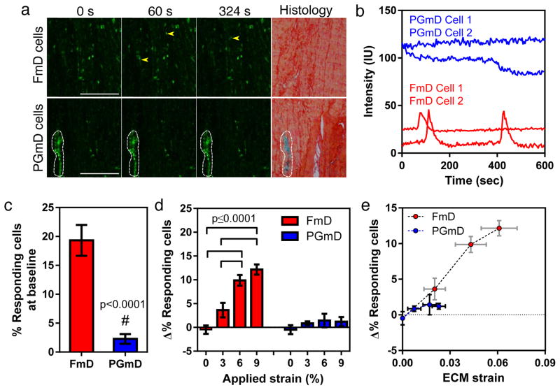
Treatment strategies to address pathologies of fibrocartilaginous tissue are in part limited by an incomplete understanding of structure-function relationships in these load-bearing tissues. There is therefore a pressing need to develop micro-engineered tissue platforms that can recreate the highly inhomogeneous tissue microstructures that are known to influence mechanotransductive processes in normal and diseased tissue. Here, we report the quantification of proteoglycan-rich microdomains in developing, ageing and diseased fibrocartilaginous tissues, and the impact of these microdomains on endogenous cell responses to physiologic deformation within a native-tissue context. We also developed a method to generate heterogeneous tissue-engineered constructs (hetTECs) with non-fibrous proteoglycan-rich microdomains engineered into the fibrous structure, and show that these hetTECs match the microstructural, micromechanical and mechanobiological benchmarks of native tissue. Our tissue-engineered platform should facilitate the study of the mechanobiology of developing, homeostatic, degenerating and regenerating fibrous tissues.


Figures

Figure 1
Figure 1. Proteoglycan-rich micro-domains (PGmDs) become more prevalent and larger in dense connective tissues with development, aging, and mechanical adaptation
Histology of (a) fetal, (b) juvenile, (c) adult bovine and (d) adult human meniscus specimens. Picrosirius Red staining indicates collagen and Alcian Blue staining indicates proteoglycans (PGs). Scale bars = 250 μm. (e) Polarized light and second harmonic generation (SHG) images of a human outer meniscus section (Alcian Blue) showing large PGmD regions lacking organized collagen. Scale bar = 250 μm. (f) Quantification of the number of PGmDs per mm2 and (g) PGmD area in individual native tissue specimens as a function of age/species. Data are presented as median ± interquartile range. Kruskal Wallis (p<0.05) with Bonferroni-Dunn’s post-hoc. p*: Bonferroni adjusted p value. (h) Correlation between median PGmD area and age in human tissue. (i) Correlation between median PGmD area and body mass index (BMI) in human tissue. Shades of gray indicate whether the specimen came from a donor that was underweight (lightest shade), of normal weight, overweight, or obese (darkest shade). Spearman’s rank correlation. Solid line: regression line; Dashed line: standard error of regression; ρs: Spearman’s rho. Blue dots indicate tissues with a diagnosed history of osteoarthritis (OA).
Figure 2
Figure 2. PGmDs attenuate local strain transmission and cell deformation in fibrocartilage
(a) Schema of mechanical test. Tissue samples were stretched uniaxially while simultaneously monitoring position and size of cells and nuclei in situ via confocal microscopy. (b) Lagrangian strain at the tissue and local ECM length scale was computed by tracking centroids of surface markers and cell nuclei, respectively. Cell strain was calculated by measuring the change in the long axis of individual cells. (c) Histology (Alcian Blue and Picrosirius Red) showing the presence of PGmDs within an imaged region and a micrograph of cell nuclei in both PGmD and FmD. Scale bar = 250 μm. Strain transfer from the tissue to ECM (FmD and PGmD) in bovine (d) fetal, (e) juvenile, (f) adult, and (g) non-OA human outer meniscus; PGmD regions showed attenuated strain transfer. ★: p<0.05 vs FmD via extra-sum-of-squares F-test; m: slope of linear regression (average strain transfer ratio). Dashed line represents 1:1 relationship. n=10–20 triads for each FmD and PGmD. (h) Finite element simulated longitudinal strain map of untreated and ChABC treated specimens at 10% applied strain. Arrow indicates PGmD. (i) PGmD/FmD strain ratio from experimental data and finite element (FE) simulations of untreated and ChABC treated specimens. (j) Digestion of proteoglycans (via ChABC treatment) from PGmDs resulted in a partial recovery of strain transfer in juvenile meniscus specimens. ★: p<0.05 vs FmD and #: p<0.05 vs PGmD via extra-sum-of-squares F-test. n= 21 triads for ChABC treated. All data are presented as mean ± s.e.m.
Figure 3
Figure 3. PGmDs alter local cell mechano-response to applied tissue level mechanical deformation
(a) Representative time series confocal snapshots at 0, 60, and 324 sec, and corresponding histology (Alcian Blue and Picrosirius Red) for a juvenile meniscus tissue. Cells in FmDs respond to 3% strain with an increase in the number of calcium oscillations over time, as indicated by the yellow arrowheads. Conversely, cells in PGmDs do not respond to stretch, but have an elevated baseline calcium signal. White outline indicates PGmD area. Scale bar = 250 μm. (b) Representative fluorescence intensity traces for cells in FmDs and PGmDs after stretch. (c) Percentage of responding cells at baseline in the FmD and PGmD regions. #: p<0.0001 vs FmD via Student’s t-test (two-tailed and unpaired). n=21 and 23 for FmD and PGmD, respectively. (d) Change in the percentage of responding cells with applied stretch relative to baseline levels. n=5–7 per strain group. One-way ANOVA (p<0.05) with Tukey’s post-hoc. (e) Percent responding cells (from Figure 3d) plotted against measured ECM strain, showing relative strain dependence. All data in (a)-(e) are presented for juvenile meniscus. All data are presented as mean ± s.e.m.
Figure 4
Figure 4. Heterogeneous tissue engineered constructs (hetTECs) reproduce the structural, compositional, and molecular features of native tissue FmDs and PGmDs
(a) Schematic of the creation of a hetTEC containing FmDs with ‘engineered-in’ PGmDs. Isolated meniscus fibrochondrocytes (MFCs) and chondrogenically differentiated mesenchymal stem cell (MSC) micro-pellets were co-seeded between two layers of aligned nanofibrous scaffold. (b) MSC micro-pellets visualized by CellTracker Red and MFCs by CellTracker Green 3 days after construct assembly. Scale bar = 200 μm. (c) Histology (Alcian Blue for PGmD and Picrosirius Red for FmD) of hetTEC after 1, 4, and 8 weeks of culture. Engineered PGmDs increase in size with culture duration. Collagen deposited by MFCs in the engineered FmD follows the underlying nanofiber direction (arrow). Scale bar = 100 μm. (d) PGmD area as a function of culture duration. Values indicate mean area at each time point. One-way ANOVA (p<0.05) with Bonferroni’s post-hoc. p*: Bonferroni adjusted p value. n=10–11 per group. Data presented as mean ± s.d. (e) Type II collagen immunostaining of hetTEC at week 8. Arrow points to engineered PGmD. Scale bar = 100 μm. (f) Type I collagen immunostaining of FmD region of hetTEC at week 8. Arrow indicates scaffold fiber direction. Scale bar = 100 μm. (g) Single cell quantitative RNA fluorescent in situ hybridization (RNA FISH) of hetTECs. Representative images showing fluorescent spots (indicated by red arrow) corresponding to individual aggrecan (AGG) mRNA in cells in either an FmD or a PGmD at week 4. Scale bar = 5 μm. (h) AGG expression (normalized to GAPDH) as measured by single cell quantitative RNA FISH. n = 152 and 250 cells for FmD and PGmD, respectively. Mann-Whitney U test (two-tailed and unpaired). Data presented as median ± interquartile range. (i) Young’s modulus (MPa) of hetTEC as a function of cellular constituents. PGmD inclusion did not change bulk tensile mechanics compared to scaffold alone. Seeding of MFCs to produce the FmD significantly increased the Young’s modulus. Dashed line represents modulus of scaffold prior to seeding (10.0 ± 0.6 MPa). +: p<0.05 vs PCL scaffold only. One-way ANOVA (p<0.05) with Bonferroni’s post-hoc. n=3 each. Data presented as mean ± s.d.
Figure 5
Figure 5. HetTECs reproduce native tissue domain-dependent strain transfer characteristics and mechano-response
(a) Color maps of local ECM strain (Exx; loading and fiber direction) for hetTECs matured for 8 weeks with application of 0, 3, 9 and 15% strain. Red box indicates location of a PGmD within the hetTEC. (b) Quantification of local ECM strain within FmD and PGmD regions of hetTEC at week 8. PGmD only: construct containing only PGmD/MSC-micro-pellets; FmD only: construct containing only FmD/seeded MFCs; PGmD/FmD: construct containing both PGmD and FmD components. #: p<0.0001 vs FmD via extra-sum-of-squares F-test; ##: p<0.0001 vs PGmD/FmD (FmD) via extra-sum-of-squares F-test; m: slope of linear regression (average strain transfer ratio). Dashed line represents 1:1 relationship. n=35~50 triads per group. (c) Representative calcium response for hetTEC cells in FmDs and PGmDs over 600 sec before or after the application of 6% strain. As in native tissue, cells in PGmDs do not respond to strain and have a higher baseline calcium level. (d) Percent increase in responding cells in hetTEC FmD and PGmD regions with application of 6% strain. #: p=0.04 vs FmD via Student’s t-test (two-tailed and unpaired). n=3 per group. All data are presented as mean ± s.e.m.
Figure 6
Figure 6. Heterogeneous tissue engineered constructs (hetTECs) match the (a) microstructural, (b) micromechanical, and (c) mechano-biological benchmarks established by heterogeneous native fibrocartilages
Box in (a) indicates range of PGmD area in native tissues. Data presented as mean ± s.d. Red and blue shaded areas in (b) indicate 99% confidence interval of linear regression of FmD and PGmD ECM native tissue strain. Red and blue dots show response in hetTEC micro-domains after 8 weeks of culture. Data presented as mean ± s.e.m. Blue and red traces in (c) show calcium signaling in PGmD and FmD regions of native and engineered hetTECs in response to applied strain. All native tissue shown are from juvenile bovine meniscus.

References

    1. Andersson GB. Epidemiological features of chronic low-back pain. Lancet. 1999;354:581–585. - PubMed
    1. Englund M, Roemer FW, Hayashi D, Crema MD, Guermazi A. Meniscus pathology, osteoarthritis and the treatment controversy. Nat Rev Rheumatol. 2012;8:412–419. - PubMed
    1. Nerurkar NL, et al. Nanofibrous biologic laminates replicate the form and function of the annulus fibrosus. Nat Mater. 2009;8:986–992. - PMC - PubMed
    1. Fisher MB, et al. Engineering meniscus structure and function via multi-layered mesenchymal stem cell-seeded nanofibrous scaffolds. J Biomech. 2015;48:1412–1419. - PMC - PubMed
    1. Baek J, et al. Meniscus tissue engineering using a novel combination of electrospun scaffolds and human meniscus cells embedded within an extracellular matrix hydrogel. J Orthop Res Off Publ Orthop Res Soc. 2015;33:572–583. - PMC - PubMed

Publication types

LinkOut - more resources