Review
. 2016 Jan;126(1):12-22.
doi: 10.1172/JCI77812. Epub 2016 Jan 4.

The pathogenesis of insulin resistance: integrating signaling pathways and substrate flux

Review

The pathogenesis of insulin resistance: integrating signaling pathways and substrate flux

Varman T Samuel et al. J Clin Invest. 2016 Jan.

Abstract

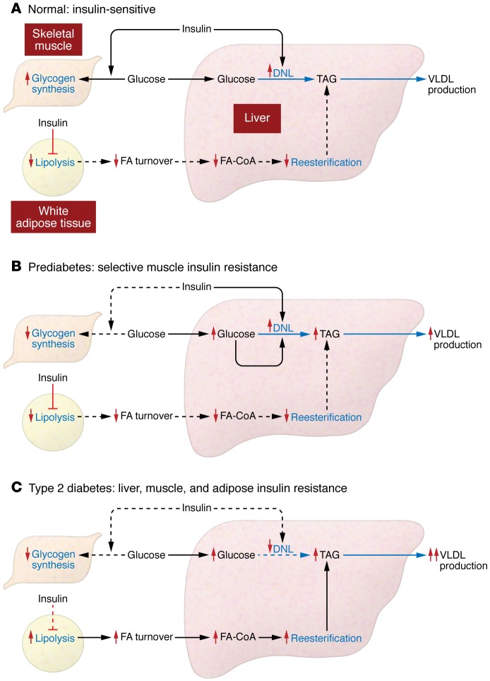
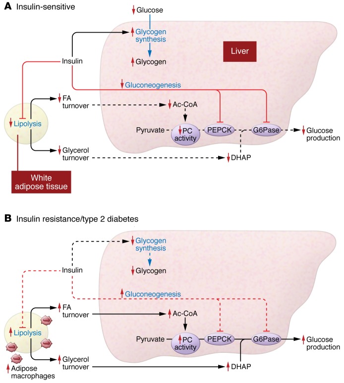
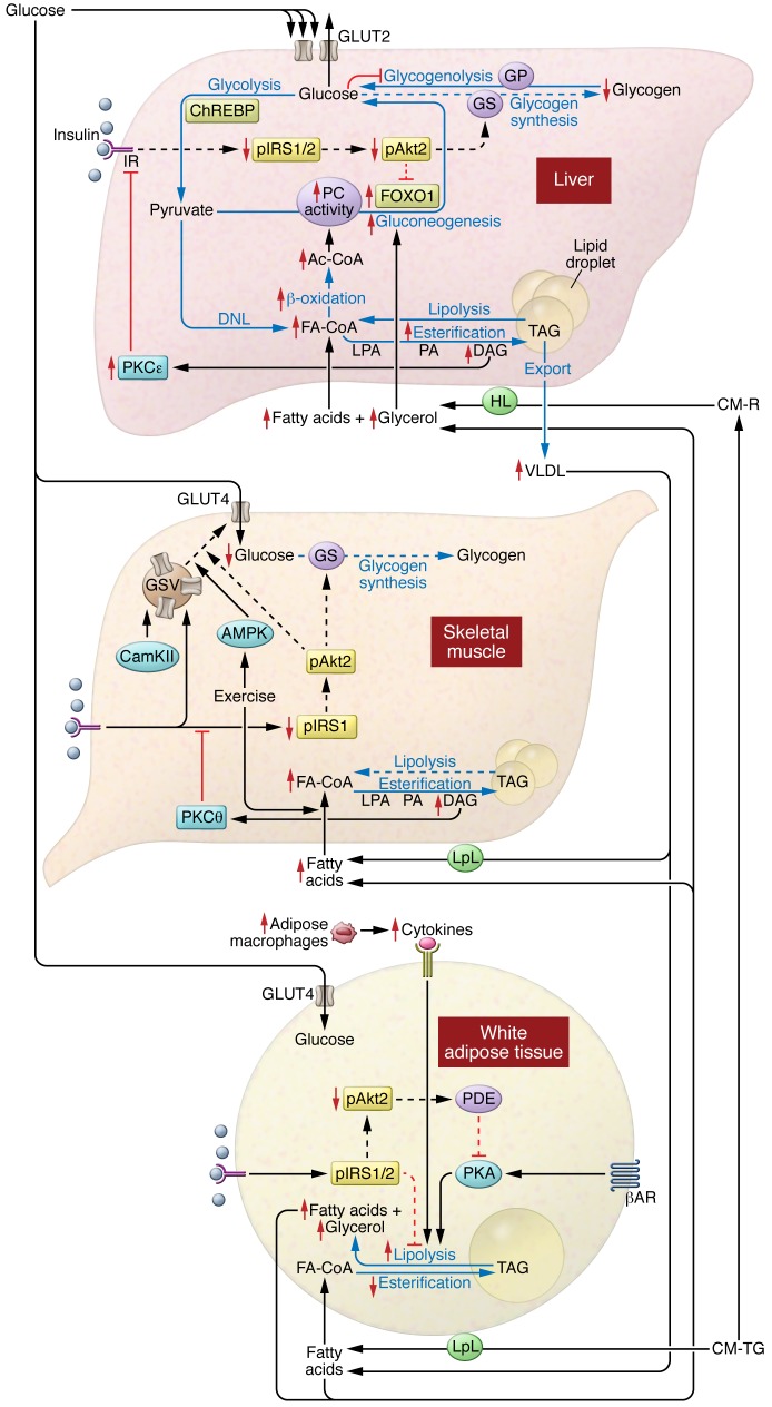
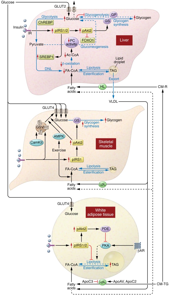
Insulin resistance arises when the nutrient storage pathways evolved to maximize efficient energy utilization are exposed to chronic energy surplus. Ectopic lipid accumulation in liver and skeletal muscle triggers pathways that impair insulin signaling, leading to reduced muscle glucose uptake and decreased hepatic glycogen synthesis. Muscle insulin resistance, due to ectopic lipid, precedes liver insulin resistance and diverts ingested glucose to the liver, resulting in increased hepatic de novo lipogenesis and hyperlipidemia. Subsequent macrophage infiltration into white adipose tissue (WAT) leads to increased lipolysis, which further increases hepatic triglyceride synthesis and hyperlipidemia due to increased fatty acid esterification. Macrophage-induced WAT lipolysis also stimulates hepatic gluconeogenesis, promoting fasting and postprandial hyperglycemia through increased fatty acid delivery to the liver, which results in increased hepatic acetyl-CoA content, a potent activator of pyruvate carboxylase, and increased glycerol conversion to glucose. These substrate-regulated processes are mostly independent of insulin signaling in the liver but are dependent on insulin signaling in WAT, which becomes defective with inflammation. Therapies that decrease ectopic lipid storage and diminish macrophage-induced WAT lipolysis will reverse the root causes of type 2 diabetes.


Figures

Figure 4
Figure 4. Insulin regulates hepatic lipid metabolism directly via hepatic insulin signaling and indirectly via adipose and muscle insulin action.
(A) Muscle insulin action promotes postprandial muscle glucose uptake, and adipose insulin action decreases hepatic fatty acid (FA) delivery and reesterification of hepatic FAs into triglycerides. Direct hepatic insulin action will activate de novo lipogenesis and conversion of excess carbohydrate substrate into triglyceride and will promote export of hepatic triglyceride to adipose tissue as very low–density lipoprotein (VLDL). DNL, de novo lipogenesis. (B) Selective muscle insulin resistance in the prediabetic state, due to selective ectopic IMCL accumulation and DAG/PKCθ-mediated inhibition of muscle insulin signaling, leads to decreased insulin-stimulated glucose transport activity. This diverts ingested glucose to the liver, where the combination of postprandial hyperinsulinemia and hyperglycemia stimulates hepatic de novo lipogenesis, resulting in increased VLDL production, hypertriglyceridemia, and reductions in plasma HDL. (C) With the progression to hepatic steatosis and impaired insulin signaling in all key insulin-responsive tissues (liver, skeletal muscle, and adipose tissue), rates of adipose tissue lipolysis are increased, resulting in increased FA delivery to liver, which results in increased hepatic esterification of fatty acids to triglyceride. This process is regulated predominately by a substrate push mechanism and is independent of insulin signaling in the hepatocyte. In contrast, hepatic de novo lipogenesis, which is dependent on hepatic insulin signaling, is reduced. Dotted lines represent decreased action or decreased flux.
Figure 3
Figure 3. Insulin regulates hepatic glucose metabolism directly via hepatic insulin action and indirectly via adipose insulin action.
(A) Insulin regulates hepatic glucose metabolism through both a direct mechanism and an indirect mechanism. The direct mechanism is mediated through activation of hepatocyte insulin receptors, which decreases hepatic glucose production acutely by activating hepatic glycogen synthesis and chronically through transcriptional downregulation of gluconeogenic enzymes, predominately by FOXO1 phosphorylation. Insulin also suppresses hepatic glucose metabolism by an indirect mechanism mediated by insulin action on WAT. Insulin inhibition of lipolysis suppresses fatty acid and glycerol turnover. This decreases fatty acid (FA) delivery to the liver, leading to reductions in hepatic acetyl-CoA (Ac-CoA) content, which in turn leads to allosteric reductions in hepatic PC activity and flux. Additionally, insulin suppression of lipolysis decreases glycerol delivery to liver and decreases conversion of glycerol to glucose. PEPCK, phosphoenolpyruvate carboxykinase; G6Pase, glucose 6-phosphatase. (B) In T2D, dysregulation of hepatic and adipose insulin action both contribute to hyperglycemia. Impaired hepatic insulin signaling, mediated by DAG/PKCε inhibition of insulin receptor kinase activity, results in reduced insulin activation of hepatic glycogen synthesis and postprandial hyperglycemia. Adipose tissue inflammation and adipose insulin resistance results in increased rates of lipolysis and increased rates of FA and glycerol delivery to the liver. Increased FA delivery to the liver increases hepatic Ac-CoA content, leading to allosteric activation of PC activity and PC flux that increases hepatic gluconeogenesis. Additionally, increased glycerol delivery to the liver further increases hepatic gluconeogenesis through a substrate push mechanism. Dotted lines represent decreased action or flux. DHAP, dihydroxyacetone phosphate.
Figure 2
Figure 2. Mechanisms of insulin resistance.
In the liver, DAG-mediated activation of PKCε impairs hepatic insulin signaling, constraining insulin-stimulated hepatic glycogen synthesis. Hepatic lipid synthesis continues unabated. In the skelatal muscle, DAG-mediated activation of PKCθ impairs muscle insulin signaling, impeding insulin-stimulated muscle glucose uptake and leading to increased glucose delivery to the liver. Exercise can still function to promote glucose uptake. In adipose tissue, cytokine release from ATMs promotes adipose lipolysis and leads to increased release of fatty acids (FAs). This will further drive hepatic lipid synthesis and activate hepatic gluconeogenesis via acetyl-CoA–mediated (Ac-CoA–mediated) activation of PC and glycerol, increasing glucose production via substrate push. IR, insulin receptor; GP; glycogen phosphorylase; GS, glycogen synthase; LPA, lysophosphatidic acid; HL, hepatic lipase; CM-R, chylomicron remnants; VLDL, very low–density lipoprotein; CAM-KII, calmodulin kinase II; IRS1/2,insulin receptor substrate 1/2; βAR, β-adrenergic receptor; CM-TG, chylomicron- triglycergides.
Figure 1
Figure 1. Insulin action promotes nutrient storage.
In the liver, nutrient flux (blue arrows) is optimized by the coordinated action of hormonal and nutrient signals. Insulin signaling through Akt2 activates glycogen synthase and decreases the transcription of gluconeogenic enzymes via inactivation of FOXO1. Insulin signaling also promotes activation and expression of SREBP1. Glucose inhibits glycogenolysis and, when metabolized, can activate ChREBP. SREBP1 and ChREBP both promote DNL. Liver uptake of fatty acids (FAs) from chylomicron remnants or FAs that spill over from peripheral lipolysis also contributes to hepatic lipid synthesis via reesterification. In skeletal muscle, insulin activates the movement of GSVs to the plasma membrane, enhancing glucose uptake and glycogen synthesis. GSV translocation can also be activated by exercise. Skeletal muscle will also take up FAs for oxidation. In adipose tissue, insulin acts to inhibit lipolysis and promote glucose uptake. Adipose tissue is the primary storage location for lipids, with a coordinate uptake of fats from chylomicrons and very low–density lipoprotein (VLDL). βAR, β-adrenergic receptor; CM-TG, chylomicron-triglycergides; IR, insulin receptor; HL, hepatic lipase; CM-R, chylomicron remnants; CAM-KII, calmodulin kinase II; GS, glycogen synthase; GP; glycogen phosphorylase; Ac-Coa, Acetyl-CoA.

Similar articles

Cited by

References

    1. Petersen KF, Laurent D, Rothman DL, Cline GW, Shulman GI. Mechanism by which glucose and insulin inhibit net hepatic glycogenolysis in humans. J Clin Invest. 1998;101(6):1203–1209. doi: 10.1172/JCI579. - DOI - PMC - PubMed
    1. Hwang JH, et al. Impaired net hepatic glycogen synthesis in insulin-dependent diabetic subjects during mixed meal ingestion. A 13C nuclear magnetic resonance spectroscopy study. J Clin Invest. 1995;95(2):783–787. doi: 10.1172/JCI117727. - DOI - PMC - PubMed
    1. Kasvinsky PJ, Shechosky S, Fletterick RJ. Synergistic regulation of phosphorylase a by glucose and caffeine. J Biol Chem. 1978;253(24):9102–9106. - PubMed
    1. Petersen KF, Laurent D, Yu C, Cline GW, Shulman GI. Stimulating effects of low-dose fructose on insulin-stimulated hepatic glycogen synthesis in humans. Diabetes. 2001;50(6):1263–1268. doi: 10.2337/diabetes.50.6.1263. - DOI - PubMed
    1. Samuel Varman T, Shulman Gerald I. Mechanisms for insulin resistance: common threads and missing links. Cell. 2012;148(5):852–871. doi: 10.1016/j.cell.2012.02.017. - DOI - PMC - PubMed

Publication types