. 2016;12(3):499-514.
doi: 10.1080/15548627.2015.1134080. Epub 2016 Jan 4.

Control of lysosomal biogenesis and Notch-dependent tissue patterning by components of the TFEB-V-ATPase axis in Drosophila melanogaster

Affiliations

Control of lysosomal biogenesis and Notch-dependent tissue patterning by components of the TFEB-V-ATPase axis in Drosophila melanogaster

Emiliana Tognon et al. Autophagy. 2016.

Abstract

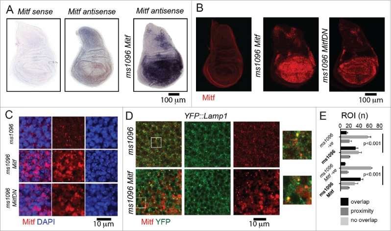
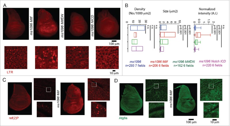
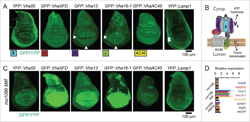
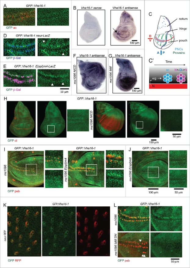
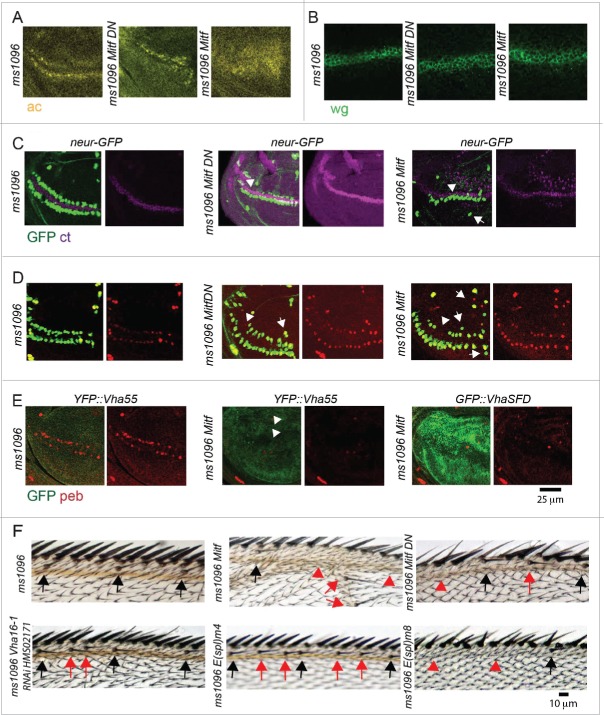
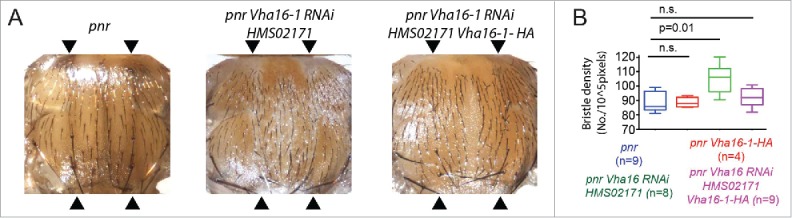
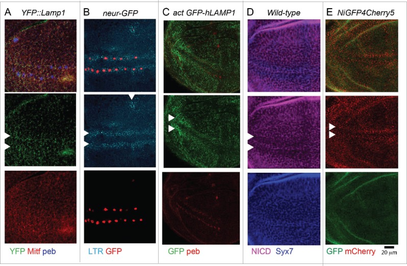
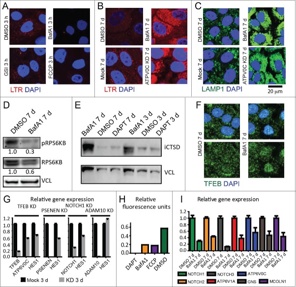
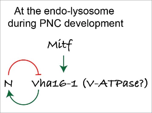
In vertebrates, TFEB (transcription factor EB) and MITF (microphthalmia-associated transcription factor) family of basic Helix-Loop-Helix (bHLH) transcription factors regulates both lysosomal function and organ development. However, it is not clear whether these 2 processes are interconnected. Here, we show that Mitf, the single TFEB and MITF ortholog in Drosophila, controls expression of vacuolar-type H(+)-ATPase pump (V-ATPase) subunits. Remarkably, we also find that expression of Vha16-1 and Vha13, encoding 2 key components of V-ATPase, is patterned in the wing imaginal disc. In particular, Vha16-1 expression follows differentiation of proneural regions of the disc. These regions, which will form sensory organs in the adult, appear to possess a distinctive endolysosomal compartment and Notch (N) localization. Modulation of Mitf activity in the disc in vivo alters endolysosomal function and disrupts proneural patterning. Similar to our findings in Drosophila, in human breast epithelial cells we observe that impairment of the Vha16-1 human ortholog ATP6V0C changes the size and function of the endolysosomal compartment and that depletion of TFEB reduces ligand-independent N signaling activity. Our data suggest that lysosomal-associated functions regulated by the TFEB-V-ATPase axis might play a conserved role in shaping cell fate.

Keywords: Notch signaling; SOP; TFEB; V-ATPase; autophagy; lysosome; mitf; patterning.


Figures

Figure 1.
Figure 1.
Mitf localization in wing imaginal discs. (A) In situ hybridization using labeled sense and antisense RNA probe for Mitf transcripts in wing discs from yellow white (control) animals and from animals overexpressing Mitf in wing disc (ms1096 Mitf). The sense probe has been used as a negative control. Dorsal is up, anterior to the left. All wing discs shown in figures are oriented as such. (B) Control wing disc and wing disc overexpressing Mitf or Mitf DN stained with anti- Drosophila Mitf antibody. (C and D) High magnifications of wing pouch tissue of the indicated genotype stained as indicated. Note that Mitf is present in the nucleus when overexpressed and in some lysosomes (close-up in D). (E) Quantification of colocalization of YFP:Lamp1 and Mitf. Colocalization across 80 regions of interest (ROI) per discs is shown as overlap. Proximity denotes nonoverlapping signal that falls within the ROI. Averages of 3 discs per genotype are graphed. -ve controls are obtained by rotating one of the 2 channels by 90 degrees.
Figure 2.
Figure 2.
Mitf regulates lysosomal biogenesis. (A) Wing discs of the indicated genotypes subjected to incorporation of LTR. High magnifications of portions of the overexpressing tissue are shown below the discs. (B) Quantification of LTR puncta density, size and normalized intensity are graphed for the indicated samples. Overexpression of Mitf or Mitf DN increases LTR incorporation in wing disc cells, while overexpression of activated NICD slightly reduces it. (C and D) Immunolocalization of ref(2)P and Atg8a in discs of the indicated genotype. Side panels are higher magnifications of the overexpressing tissue with insets shown below them.
Figure 3.
Figure 3.
Mitf controls transcription of V-ATPase genes. (A) Pattern of expression in wing discs of the tagged lines for the indicated subset of Drosophila orthologs of TFEB target genes. Arrowheads point to areas with distinctive pattern of expression. (B) Schematics of the structure and function of V-ATPase with positioning of the subunits analyzed. (C) Pattern of expression in wing discs of the tagged lines for the indicated subset of Drosophila orthologs of TFEB target genes upon overexpression of Mitf. Note that V-ATPase subunit expression but not Lamp1 expression is controlled by Mitf. (D) Transcript levels of the indicated putative Mitf targets in wing discs by Q-PCR. RpL32 has been used as housekeeping control. The values represents means ± s.d of 2 independent experiments.
Figure 4.
Figure 4.
Vha16-1 expression is elevated in SOPs. (A) A high magnification of the anterior part of the wing pouch of wing discs of the indicated genotypes stained as indicated. Note that GFP::Vha16-1 expression is elevated in ac-expressing tissue. (B) In situ hybridization using labeled sense and antisense RNA probes for Vha16-1 transcripts in control wing discs. Note the high expression in 2 stripes of tissue abutting the anterior D/V boundary (arrow) when using the antisense probe. (C) Schematic representation of the patterning of a larval wing disc. The patterning features relevant for this study are indicated. Dl, N ligand Delta. (D and E) A high magnification of the anterior part of the wing pouch of wing discs of the indicated genotypes stained as indicated. Note that GFP::Vha16-1 expression is elevated in neur-LacZ-positive cell (D, arrowheads) and low in E(spl)m4 -positive cells (E, arrowheads). Arrowheads in E indicate examples of SOP cells that are not E(spl)m4-positive and have high GFP::Vha16-1 expression. (F and G) In situ hybridization using an antisense RNA probe for Vha16-1 transcripts of discs overexpressing NICD or Mitf. Note the very different transcriptional modulation of Vha16-1 expression upon overexpression. (H) Discs of the indicated genotype stained to detect the N target cut (ct). Note that GFP::Vha16-1 expression is very low and not patterned in ct -positive tissue. Insets enlarging corresponding areas of the pouch are show on the sides of each disc. (I and J) GFP::Vha16-1 wing disc overexpressing the N target E(Spl) genes m8, and m4 under ms1096-Gal4. Note that overexpression of E(Spl)m8 results in loss of sensory organs and of GFP::Vha16-1 expression at the anterior margin, while overexpression of E(Spl)m4 leads to formation of ectopic SOPs expressing GFP::Vha16-1. Insets enlarging corresponding areas of the pouch are show on the sides of each disc. (K) Pupal nota of the indicated genotype dissected 20 h after puparium formation. Note that elevated GFP::Vha16-1 expression is maintained along the SOP lineage. (L) High magnifications of the anterior part of the wing pouch of wing discs of the indicated genotypes stained as indicated. The image is a maximal projection of several sections. Note that overexpression of Mitf DN disrupts the pattern of GFP::Vha16-1 expression in SOPs and leads to missing and ectopic SOPs (arrowheads).
Figure 5.
Figure 5.
Misexpression of Mitf perturbs SOP development. (A and D) High magnification of the anterior part of the wing disc pouch of the indicated genotype stained as indicated. Note that Mitf and Mitf DN overexpression results in perturbation of the expression of ac protein (A), no perturbation of wg or ct expression at the D/V boundary (B and C), formation of misplaced or ectopic neur-GFP- ct -, (C) and neur-GFP- peb - (D) positive cells. Some of the ectopic ct and peb -positive cells are negative for neur-GFP and could represent incomplete SOP commitment. (E) Presence of normal and ectopic peb -positive cells is reduced to different extent in YFP::Vha55 discs overexpressing Mitf, when compared to YFP::GFP discs not overexpressing Mitf. A similar lack of peb -positive cells is observed in GFP::VhaSFD discs overexpressing Mitf. Note that the genetic null tagged forms of these genes are highly expressed, due to induction by Mitf. (F) High magnification of the antero-distal dorsal area of the margin of adult wings of the indicated genotypes. The stereotypic position of sensory margin bristles is shown by black arrows. Expression of the indicated constructs in wing discs results in loss (red arrowheads) or misplacement of sensory bristles (red arrows).
Figure 6.
Figure 6.
Vha16-1 is required for in pupal SOPs differentiation. (A) Phenotypic defects associated with RNAi-mediated knockdown of Vha16-1 expression with the indicated RNAi line using the driver pannier-Gal4 (pnr) compared to control. Defects are rescued by concomitant expression of the RNAi lines and a RNAi-resistant Vha16-1-HA construct. The domain of pannier-Gal4 expression is delimited by arrowheads. (B) Quantification of the number of bristle/area (bristle density) relative to the experiment as in A. Statistical analysis is based on Kruskal Wallis Test with Dunn multiple comparison relative to control.
Figure 7.
Figure 7.
The endolysosomal system at sites of PNC development is distinctive. (A and E) High magnification of the anterior part of the wing pouch of wing discs of the indicated genotypes, stained as indicated. Arrowheads point to the approximate location of PNC. Note that compared to surrounding epithelial cells, PNC cells show a slightly higher amount of YFP::Lamp1-positive lysosomes (A), a higher number of acidified organelles (B) and of GFP-hLAMP1 puncta (C), overall less N (D) and more endolysosomal N (E).
Figure 8.
Figure 8.
The TFEB-V-ATPase axis regulates lysosomal function and basal N signaling in human cells. (A) LTR assay indicates that acute treatment with both BafA1 and FCCP, which dissipates lysosomal pH independently of V-ATPase, impairs acidification in MCF10A cells. (B and C) Chronic inhibition of V-ATPase or KD of ATP6V0C in contrast causes an expansion of the acidified (B) and LAMP1-positive lysosomal organelles (C). (D) Relative to the control, western blot analysis of MTOR signaling of lysate from MCF10A cells treated as indicated reveals a reduction in the level of phosphorylated RPS6KB (pS6K), a measure of active MTOR signaling. (E) Western blot analysis of MCF10A lysates treated as indicated shows that chronic inhibition of V-ATPase leads to accumulation of the 52 and 44 KDa immature CTSD forms (iCTSD), a sign of impaired lysosomal function. (F) Chronic V-ATPase inhibition results in high levels of nuclear TFEB compared to control. (G) siRNA against TFEB reduce the level of TFEB, ATP6V0C and HES1 mRNA expression compared to control. A similar reduction in the level of N signaling is observed after knocking down the expression of the γ-secretase component, PSENEN or of the NOTCH1 receptor. In contrast, efficient knockdown of ADAM10, which is dispensable for ligand-independent signaling, does not reduce HES1 levels. (H) Chronic treatment with both BafA1 and FCCP compromises γ-secretase substrate cleavage. (I) Chronic inhibition of the V-ATPase reduces the levels of expression of N receptors among other genes.
Figure 9.
Figure 9.
Proposed role of lysosomes, and of the TFEB-V-ATPase axis in patterning. A model for the activity of Mitf and V-ATPase in PNC regions during Drosophila wing disc development. [Change to “N” or “N/Notch.”]

References

    1. Forgac M. Vacuolar ATPases: rotary proton pumps in physiology and pathophysiology. Nat Rev Mol Cell Biol 2007; 8:917-29; PMID:17912264; http://dx.doi.org/ 10.1038/nrm2272 - DOI - PubMed
    1. Hinton A, Bond S, Forgac M. V-ATPase functions in normal and disease processes. Pflugers Arch 2009; 457:589-98; PMID:18026982; http://dx.doi.org/ 10.1007/s00424-007-0382-4 - DOI - PubMed
    1. Buechling T, Bartscherer K, Ohkawara B, Chaudhary V, Spirohn K, Niehrs C, Boutros M. Wnt/Frizzled signaling requires dPRR, the Drosophila homolog of the prorenin receptor. Curr Biol 2010; 20:1263-8; PMID:20579883; http://dx.doi.org/ 10.1016/j.cub.2010.05.028 - DOI - PubMed
    1. Cruciat C-M, Ohkawara B, Acebron SP, Karaulanov E, Reinhard C, Ingelfinger D, Boutros M, Niehrs C. Requirement of prorenin receptor and vacuolar H+-ATPase-mediated acidification for Wnt signaling. Science 2010; 327:459-63; PMID:20093472; http://dx.doi.org/ 10.1126/science.1179802 - DOI - PubMed
    1. Gleixner EM, Canaud G, Hermle T, Guida MC, Kretz O, Helmstädter M, Huber TB, Eimer S, Terzi F, Simons M. V-ATPase/mTOR signaling regulates megalin-mediated apical endocytosis. Cell Rep 2014; 8:10-9; PMID:24953654; http://dx.doi.org/ 10.1016/j.celrep.2014.05.035 - DOI - PubMed

Publication types

MeSH terms