. 2016;12(2):410-23.
doi: 10.1080/15548627.2015.1132134.

Neuroprotection by selective neuronal deletion of Atg7 in neonatal brain injury

Affiliations

Neuroprotection by selective neuronal deletion of Atg7 in neonatal brain injury

Cuicui Xie et al. Autophagy. 2016.

Abstract

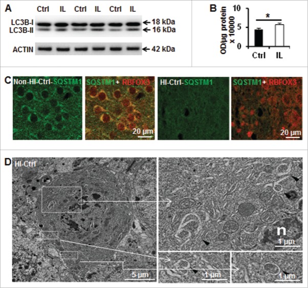
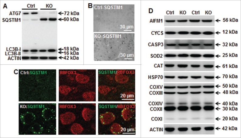
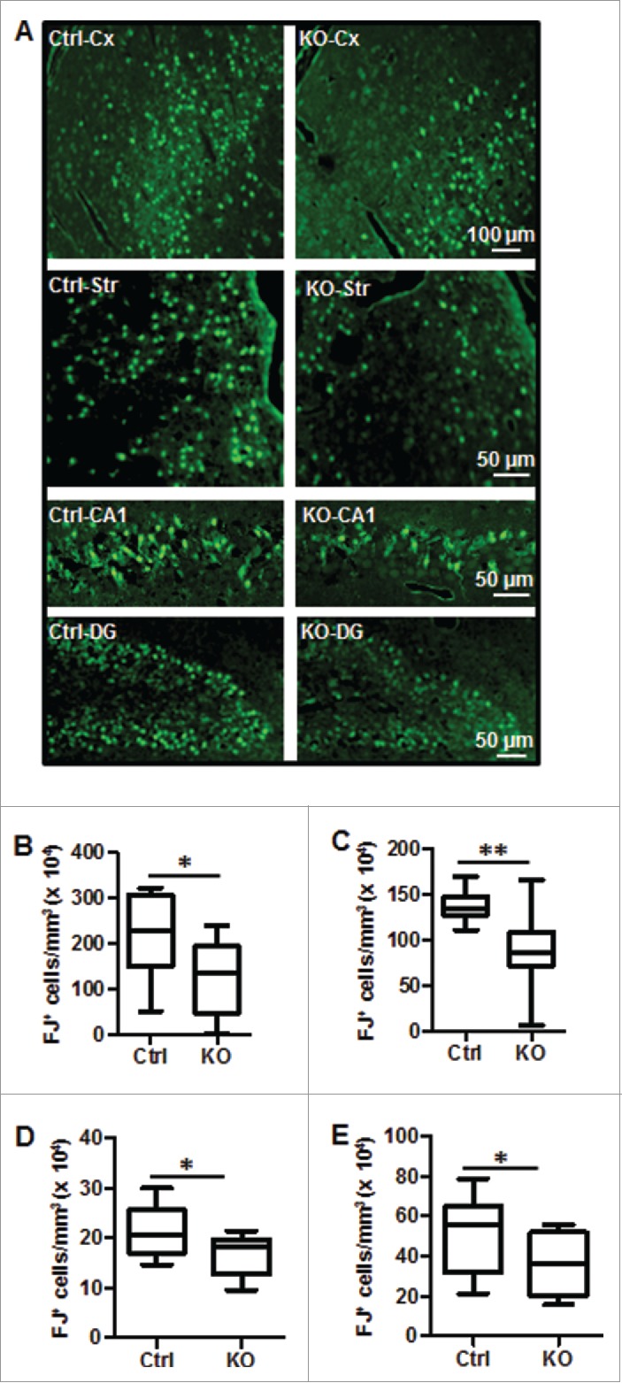
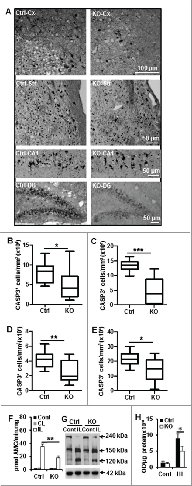
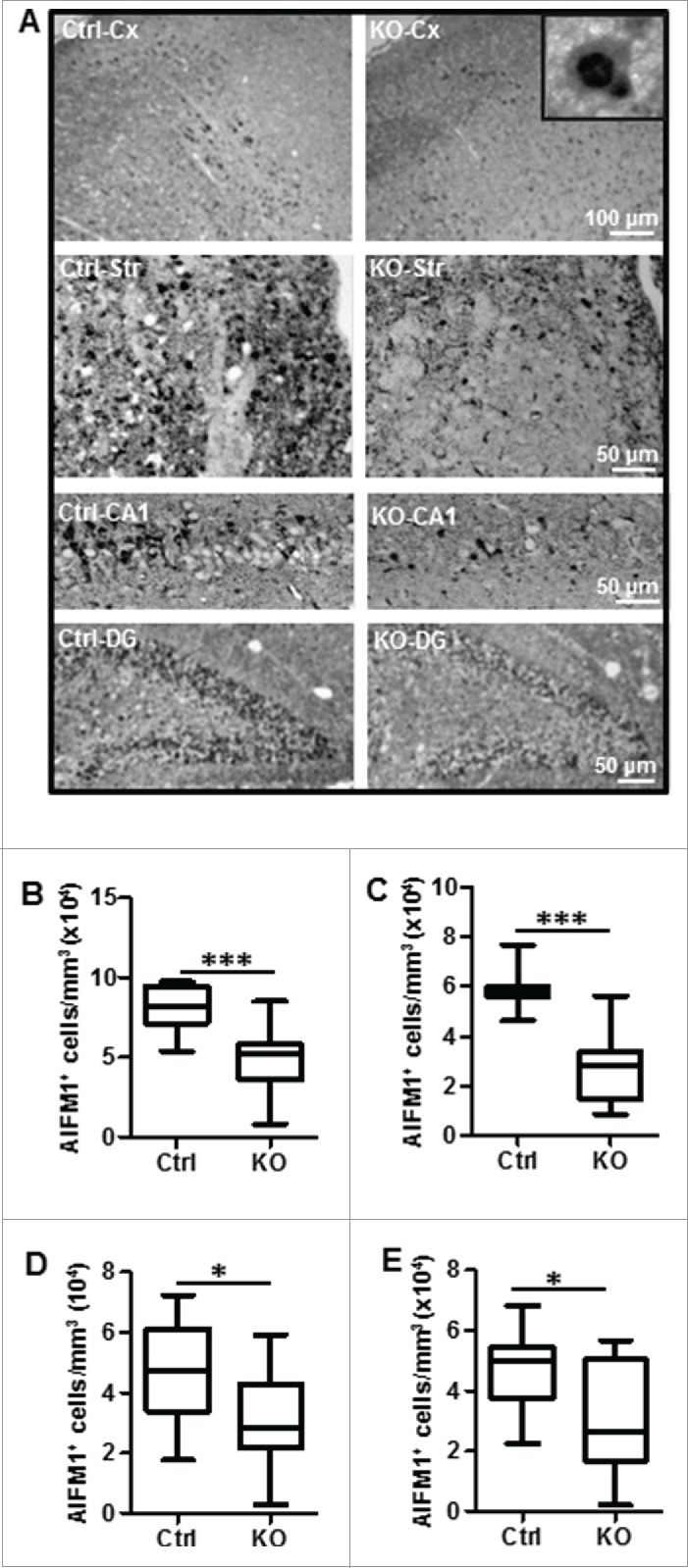
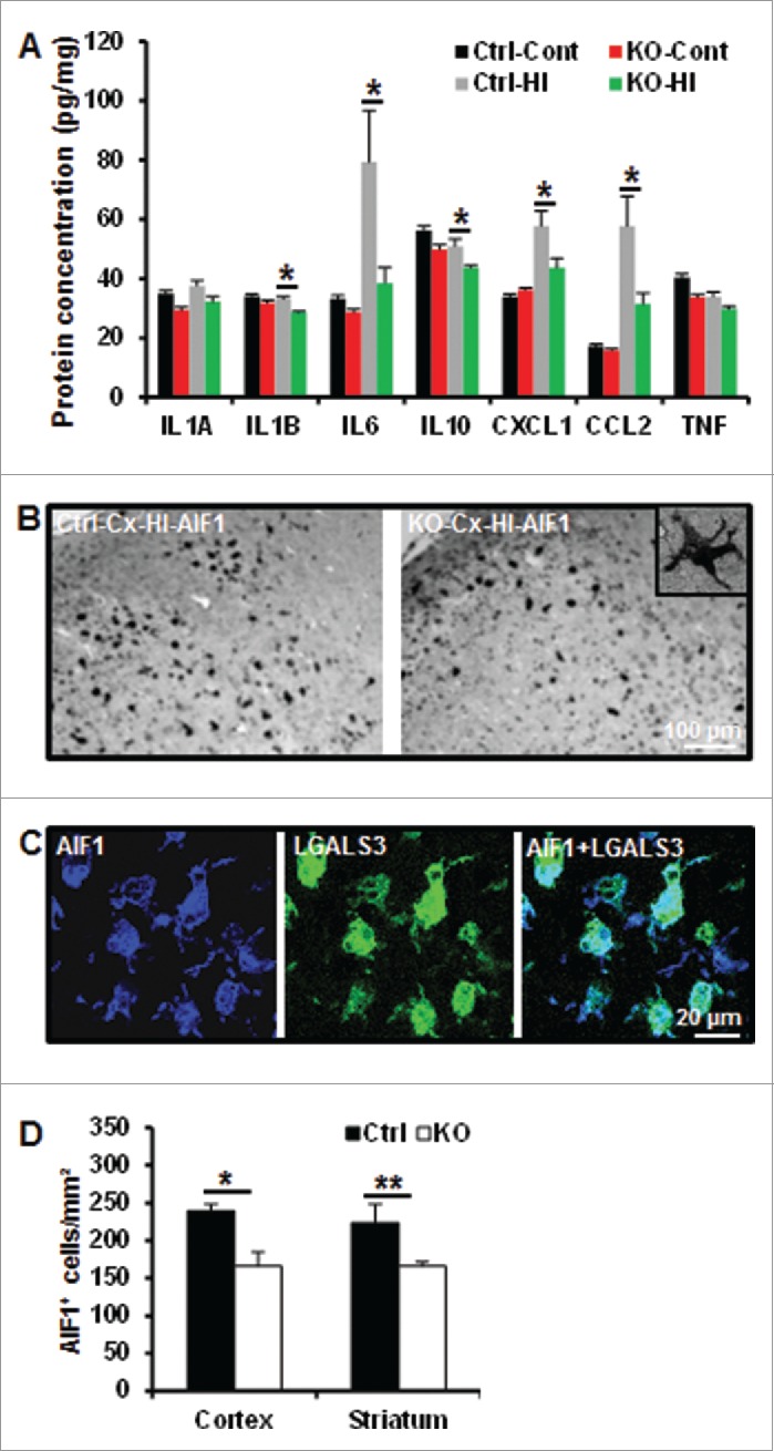
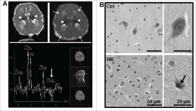
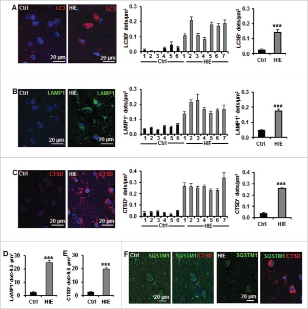
Perinatal asphyxia induces neuronal cell death and brain injury, and is often associated with irreversible neurological deficits in children. There is an urgent need to elucidate the neuronal death mechanisms occurring after neonatal hypoxia-ischemia (HI). We here investigated the selective neuronal deletion of the Atg7 (autophagy related 7) gene on neuronal cell death and brain injury in a mouse model of severe neonatal hypoxia-ischemia. Neuronal deletion of Atg7 prevented HI-induced autophagy, resulted in 42% decrease of tissue loss compared to wild-type mice after the insult, and reduced cell death in multiple brain regions, including apoptosis, as shown by decreased caspase-dependent and -independent cell death. Moreover, we investigated the lentiform nucleus of human newborns who died after severe perinatal asphyxia and found increased neuronal autophagy after severe hypoxic-ischemic encephalopathy compared to control uninjured brains, as indicated by the numbers of MAP1LC3B/LC3B (microtubule-associated protein 1 light chain 3)-, LAMP1 (lysosomal-associated membrane protein 1)-, and CTSD (cathepsin D)-positive cells. These findings reveal that selective neuronal deletion of Atg7 is strongly protective against neuronal death and overall brain injury occurring after HI and suggest that inhibition of HI-enhanced autophagy should be considered as a potential therapeutic target for the treatment of human newborns developing severe hypoxic-ischemic encephalopathy.

Keywords: ATG7; apoptosis; autophagy; caspase; hypoxic-ischemic encephalopathy; newborn.


Figures

Figure 1.
Figure 1.
Hypoxia-ischemia induces neuronal autophagy in neonatal Atg7flox/+; Nes-Cre mice. (A) Representative immunoblotting of LC3B in Ctrl (Atg7flox/+; Nes-Cre) mice and (B) its corresponding quantification showed that LC3B-II (16 kDa) was increased in the ipsilateral hemisphere (IL) 24 h after HI compared to an uninjured control brain, (*, P < 0.05, n = 6). (C) Immunostaining of SQSTM1 in the cortex of Ctrl mice suggesting a decrease of its staining in neurons (RBFOX3) 24 h after HI. (D) Dying neurons in Ctrl mice exhibited increased electron density and nuclei with chromatin condensation. They possessed also typical autophagosomes with double membranes in the perikarya (black arrowheads) 24 h after HI. Squared areas are enlarged in the right panels.
Figure 2.
Figure 2.
Neuronal Atg7 deficiency prevents neuronal basal autophagy and does not alter expression of mitochondria and cell death-related proteins. (A) Representative immunoblotting of ATG7, SQSTM1, and LC3BB in the atg7 KO (Atg7flox/flox; Nes-Cre) and Ctrl (Atg7flox/+; Nes-Cre) mice showing that ATG7 was strongly decreased in KO mice leading to an accumulation of SQSTM1 and absence of LC3BB-II. (B) Representative immunostaining of SQSTM1 in the cortex of Ctrl and atg7 KO mice in the non-HI control condition, confirming the accumulation of SQSTM1 in KO compared to the faint and diffuse expression in Ctrl. (C) Punctate and strong SQSTM1 staining was observed in the cytoplasm of neuronal cells (RBFOX3) in the cortex of atg7 KO mice, but not in the Ctrl as shown by RBFOX3 and SQSTM1. (D) Representative immunoblots from cortical tissue homogenates of P9 Ctrl and atg7 KO mice. Quantification did not show any significant differences between the 2 types of mice for cell death-related proteins AIFM1, CYCS and CASP3, mitochondria-related proteins (SOD2, HSP70, CAT and mitochondrial respiratory chain complexes (COXV, COXIII, COXIV, COXII, COXI). (n = 6/group). KO: atg7 KO (Atg7flox/flox; Nes-Cre) and Ctrl: Atg7flox/+; Nes-Cre.
Figure 3.
Figure 3.
Neuronal Atg7 deficiency reduced hypoxia-ischemia-induced neuronal autophagy and brain injury. (A) Representative immunoblots of LC3BB from non-HI control (Cont) and HI ipsilateral (IL) hemispheres of Ctrl and atg7 KO mice 24 h after HI and the corresponding quantification of LC3BB-II showed that Atg7 deletion completely prevented HI-induced LC3BB-II increase, (*P < 0.05, n = 6/group). (B) Representative MAP2 staining in the ipsilateral hemisphere of the hippocampus (left) and striatum (right) level with the indication of MAP2 negative areas. (C) Representative MAP2 staining of coronal brain sections 8 d after HI at the levels of the dorsal hippocampus (left panels) and striatum (right panels) from Ctrl and atg7 KO mice and (D) measure of total tissue loss volume demonstrated a strong reduction of the lesion in the atg7 KO mice (Ctrl: n = 38, atg7 KO: n = 28), (**, P < 0.01). (E) Pathological scores performed in the cortex (Cx), hippocampus (Hip), striatum (Str), and thalamus (Tha) confirmed the greater resistance of atg7 KO mice compared to Ctrl mice, **, P < 0.01. (F) There was no difference in the neuroprotection provided by Atg7 deletion between males (Ctrl: n = 22, atg7 KO: n = 16) and females (Ctrl: n = 16, atg7 KO: n = 12) (*, P < 0.05). (G) The neuronal cell architecture in the Ctrl and atg7 KO mice in non-HI control and 24 h after HI indicated that dying neurons with condensed chromatin were more frequent in Ctrl than in atg7 KO. Squared areas are enlarged in the right panels. Moreover, the neuronal architecture was relatively well preserved in the atg7 KO mouse compared to that of the Ctrl mice. KO: atg7 KO (Atg7flox/flox; Nes-Cre) and Ctrl: Atg7flox/+; Nes-Cre.
Figure 4.
Figure 4.
Neuronal Atg7 deficiency reduced hypoxia-ischemia-induced neuronal cell death. (A) Representative Fluoro-Jade (FJ) staining 24 h after HI and the corresponding quantifications of the number of FJ-positive cells (B) in the border zone of the cortical infarction (Cx) (2,093,000 ± 287,600/mm3 vs. 1,259,000 ± 230,200/mm3; *, P<0.05), (C) in the striatum (Str) (1,368,000 ± 46,860/mm3 vs. 912,300 ± 134,100/mm3; **, P < 0.01), (D) in the entire CA1 (209,900 cells ± 14,600 cells/mm3 vs. 167,500 cells ± 12,310 cells/mm3; *, P < 0.05) and (E) in the dentate gyrus (DG) (500,700 cells ± 56,880 cells/mm3 vs. 344,000 cells ± 45,150 cells/mm3; *, P < 0.05, n = 11/group). KO: atg7 KO (Atg7flox/flox; Nes-Cre) and Ctrl: Atg7flox/+; Nes-Cre.
Figure 5.
Figure 5.
Neuronal Atg7 deficiency decreased CASP3 activation after hypoxia-ischemia. (A) Representative active CASP3/caspase-3 staining 24 h after HI and the corresponding quantifications of the number of CASP3-positive cells (B) in the cortex (Cx) (81,630 ± 7,671/mm3 vs. 51,210 ± 11,970/mm3; *, P < 0.05), (C) in the striatum (Str) (132,800 ± 5,368/mm3 vs. 47,350 ± 13,110/mm3; ***, P < 0.001), (D) in the CA1 (41,810 cells ± 3,243 cells/mm3 vs. 22,860 cells ± 4,685 cells/mm3; **, P < 0.01) and (E) dentate gyrus (DG) (214,000 cells ± 14,700 cells/mm3 vs. 144,000 cells ± 23,940 cells/mm3; *, P < 0.05, n = 11/group). (F) CASP3 enzymatic activity 24 h after HI was 50% lower in the ipsilateral (IL) hemisphere of atg7 KO mice than in Ctrl (n = 10 for Ctrl, n = 6 for atg7 KO). There was no difference in the nonischemic control brains (Cont) (n = 13 for Ctrl and n = 6 for atg7 KO) or in the contralateral (CL) hemispheres (n = 10 for Ctrl, n = 6 for atg7 KO). (G) Representative SPTAN1/α-fodrin (240 kDa) western blots from control (Cont) and ipsilateral (IL) hemispheres 24 h after HI and (H) the quantification of the caspase-dependent 120 kDa breakdown products confirmed that CASP3 activation was more pronounced in the Ctrl mice. (*, P < 0.05, n = 6/group). KO: atg7 KO (Atg7flox/flox; Nes-Cre) and Ctrl: Atg7flox/+; Nes-Cre.
Figure 6.
Figure 6.
Neuronal Atg7 deficiency reduced hypoxia-ischemia-induced AIFM1 nuclear translocation. (A) Representative AIFM1 staining 24 h after HI and the corresponding quantifications of the number of AIFM1-positive nuclei (B) in the cortex (Cx) (80,610 nuclei ± 4,038 nuclei/mm3 vs. 49,060 nuclei ± 6,115 nuclei/mm3; ***, P < 0.001), (C) in the striatum (Str) (59,610 nuclei ± 2,322 nuclei/mm3 vs. 28,020 nuclei ± 4,287 nuclei/mm3; ***, P < 0.001), (D) in the CA1 (45,620 nuclei ± 3,849 nuclei/mm3 vs. 30,350 nuclei ± 5,123 nuclei/mm3; *, P < 0.05) and (E) dentate gyrus (DG) (48,260 nuclei ± 5,278 nuclei/mm3 vs. 30,730 nuclei ± 4,294 nuclei/mm3; *, P < 0.05). n = 11/group. KO: atg7 KO (Atg7flox/flox; Nes-Cre) and Ctrl: Atg7flox/+; Nes-Cre.
Figure 7.
Figure 7.
Neuronal Atg7 deficiency attenuated hypoxia-ischemia-induced cytokine and chemokine expression and microglial activation. (A) Luminex assay of the cytosolic fraction of nonischemic (Cont) (n = 9 for Ctrl and n = 7 for atg7 KO) and for hypoxic-ischemic cortical tissue 24 h after HI (n = 9 for Ctrl and n = 7 for atg7 KO) demonstrated a decreased IL6, CXCL1, CCL2 and IL1B in KO mice. (B) Representative AIF1 immunostaining in the cortex of both Ctrl and atg7 KO mice 24 h after HI. (C) Double labeling of AIF1 and activated microglia marker galectin-3 (LGALS3) in the HI cortex. (D) Quantification of the number of AIF1-positive cells in both the cortex (*, P<0.05) and the striatum (**, P<0.01) confirmed a less pronounced microglia activation in KO mice. (n = 11/group). KO: atg7 KO (Atg7flox/flox; Nes-Cre) and Ctrl: Atg7flox/+; Nes-Cre.
Figure 8.
Figure 8.
Lentiform nucleus of asphyxiated human term newborns is highly vulnerable. (A) Magnetic resonance (MR) diffusion-weighted images with ADC maps (top panel) of hypoxic-ischemic encephalopathy (HIE) case 1 (left) at 36 h of life and HIE case 7 (right) at 23 h of life showed bilateral restricted diffusion in the basal ganglia (mainly in the putamen, arrowheads) and thalamus (long arrows) as well as in the cortex of case 7 (dashed arrows). MR multivoxel proton spectroscopy of HIE case 7 showing a negative double lactate peak at 1.2 ppm indicating an acute energy failure in this region (white arrow) at 23 h of life (lower panel). (B) Representative hematoxylin-eosin staining of the lentiform nucleus region showed the presence of numerous dying neurons (cell shrinkage and pyknotic nuclei, arrow) in newborns subjected to perinatal asphyxia but not in controls (Ctrl).
Figure 9.
Figure 9.
Neuronal autophagy is enhanced in the lentiform nucleus of asphyxiated human term newborns. (A) Representative confocal images of LC3B staining (red) in neurons in the lentiform nuclei of human newborns, and quantifications of the numbers of LC3B-positive dots per neuron per μm2 (left histogram) in 6 controls (Ctrl) and 7 hypoxic-ischemic encephalopathy (HIE) cases, showing an increase in autophagosome in all HIE cases. Right histogram showed the average numbers of LC3B-positive dots of control (0.021 ± 0.007) and HIE (0.149 ± 0.018) cases. Representative confocal images of (B) LAMP1 staining (green) and (C) CTSD staining (red) in lentiform nuclei of human newborns and the quantifications of the numbers of positive dots per neuron per μm2 (left histograms) in the different cases individually demonstrated an increase in lysosomes in all HIE cases. Right histograms showed the average numbers of positive dots of control (LAMP1: 0.046 ± 0.005; CTSD: 0.035 ± 0.004) and HIE (LAMP1: 0.172 ± 0.013; CTSD: 0.260 ± 0.014). Quantifications showing the average percentages of (D) LAMP1- and (E) CTSD-positive dots larger than 0.5 μm2 per neuron in control (LAMP1: 2.5 ± 1.9%; CTSD: 2.6 ± 1.8%) and HIE cases (LAMP1: 24.8 ± 6.2%; CTSD: 19.8 ± 3.6%). (F) Representative confocal images of SQSTM1 staining (green) and CTSD (red) in neurons in human newborn lentiform nuclei showed that SQSTM1 expression was not increased (or accumulated) in neurons of HIE cases (right panels) (***, P<0.001).

References

    1. Mwaniki MK, Atieno M, Lawn JE, Newton CR. Long-term neurodevelopmental outcomes after intrauterine and neonatal insults: a systematic review. Lancet 2012; 379:445-52; PMID:22244654; http://dx.doi.org/10.1016/S0140-6736(11)61577-8 - DOI - PMC - PubMed
    1. Krageloh-Mann I, Helber A, Mader I, Staudt M, Wolff M, Groenendaal F, DeVries L. Bilateral lesions of thalamus and basal ganglia: origin and outcome. Dev Med Child Neurol 2002; 44:477-84; PMID:12162385; http://dx.doi.org/10.1111/j.1469-8749.2002.tb00309.x - DOI - PubMed
    1. Northington FJ, Chavez-Valdez R, Martin LJ. Neuronal cell death in neonatal hypoxia-ischemia. Ann Neurol 2011; 69:743-58; PMID:21520238; http://dx.doi.org/10.1002/ana.22419 - DOI - PMC - PubMed
    1. Carlsson Y, Schwendimann L, Vontell R, Rousset CI, Wang X, Lebon S, Charriaut-Marlangue C, Supramaniam V, Hagberg H, Gressens P, et al.. Genetic inhibition of caspase-2 reduces hypoxic-ischemic and excitotoxic neonatal brain injury. Ann Neurol 2011; 70:781-9; PMID:21674587; http://dx.doi.org/10.1002/ana.22431 - DOI - PubMed
    1. Zhu C, Wang X, Deinum J, Huang Z, Gao J, Modjtahedi N, Neagu MR, Nilsson M, Eriksson PS, Hagberg H, et al.. Cyclophilin A participates in the nuclear translocation of apoptosis-inducing factor in neurons after cerebral hypoxia-ischemia. J Exp Med 2007; 204:1741-8; PMID:17635954; http://dx.doi.org/10.1084/jem.20070193 - DOI - PMC - PubMed

Publication types

MeSH terms