. 2016 Jan 4;11(1):e0146279.
doi: 10.1371/journal.pone.0146279. eCollection 2016.

The Role of Indoleamine 2,3-Dioxygenase in Diethylnitrosamine-Induced Liver Carcinogenesis

Affiliations

The Role of Indoleamine 2,3-Dioxygenase in Diethylnitrosamine-Induced Liver Carcinogenesis

Yuhei Shibata et al. PLoS One. .

Abstract

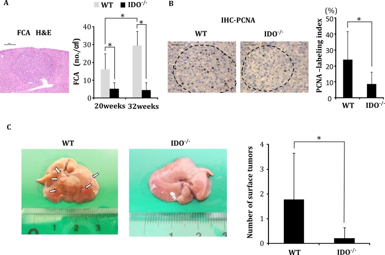
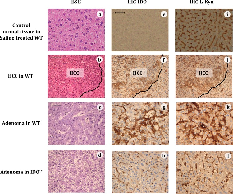
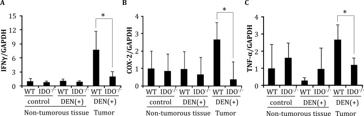
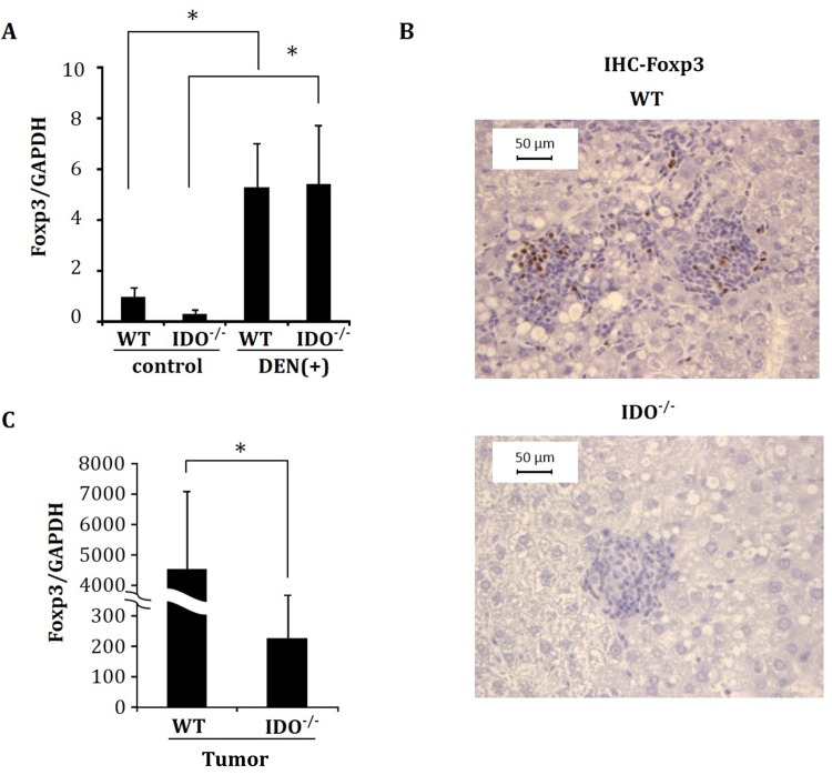
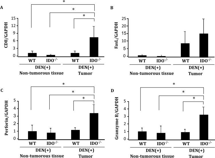
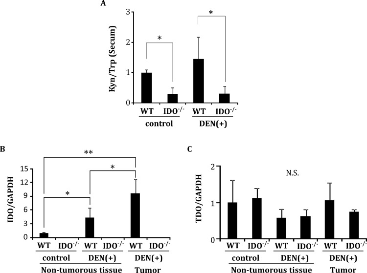
Indoleamine 2,3-dioxygenase (IDO), a tryptophan-catabolizing intracellular enzyme of the L-kynurenine pathway, causes preneoplastic cells and tumor cells to escape the immune system by inducing immune tolerance; this mechanism might be associated with the development and progression of human malignancies. In the present study, we investigated the role of IDO in diethylnitrosamine (DEN)-induced hepatocarcinogenesis by using IDO-knockout (KO) mice. To induce hepatocellular carcinoma (HCC), hepatic adenoma, and preneoplastic hepatocellular lesions termed foci of cellular alteration (FCA), male IDO-wild-type (WT) and IDO-KO mice with a C57BL/6J background received a single intraperitoneal injection of DEN at 2 weeks of age. The mice were sacrificed to evaluate the development of FCA and hepatocellular neoplasms. HCC overexpressed IDO and L-kynurenine compared to surrounding normal tissue in the DEN-treated IDO-WT mice. The number and cell proliferative activity of FCAs, and the incidence and multiplicity of HCC were significantly greater in the IDO-WT than in the IDO-KO mice. The expression levels of the IDO protein, of L-kynurenine, and of IFN-γ, COX-2, TNF-α, and Foxp3 mRNA were also significantly increased in the DEN-induced hepatic tumors that developed in the IDO-WT mice. The mRNA expression levels of CD8, perforin and granzyme B were markedly increased in hepatic tumors developed in IDO-KO mice. Moreover, Foxp3-positive inflammatory cells had infiltrated into the livers of DEN-treated IDO-WT mice, whereas fewer cells had infiltrated into the livers of IDO-KO mice. Induction of IDO and elevation of L-kynurenine might play a critical role in both the early and late phase of liver carcinogenesis. Our findings suggest that inhibition of IDO might offer a promising strategy for the prevention of liver cancer.


Conflict of interest statement

Competing Interests: The authors have declared that no competing interests exist.

Figures

Fig 1
Fig 1. Analysis of pre-neoplastic lesions and nodules of DEN-treated mice.
Development of foci of cellular alteration (FCA), the proliferating cell nuclear antigen (PCNA)-labeling indices of the FCA, and the nodule frequency on the liver surface of DEN-treated experimental mice. (A) A representative photograph of a FCA that developed in DEN-treated WT mice at 32 weeks of age (H&E staining, left panel) and the average number of FCA in DEN-treated WT and IDO-/- mice at 20 and 32 weeks of age (right panel). (B) Representative photographs of PCNA-immunohistochemical analysis of the FCA that developed in DEN-treated WT mice and IDO-/- mice at 32 weeks of age (left panels). The PCNA-labeling indices of the FCA that developed in the experimental mice were determined by counting the number of PCNA-positive nuclei in the FCA (right panel). (C) Tumor nodules on the liver surface of DEN-treated WT and IDO-/- mice at 32 weeks. The white arrows indicate tumor nodules (left panel). The number of liver surface tumors in IDO-/- mice was significantly lower than that in wild-type mice (right panel). *, P<0.01 Data points represent the mean ± SD.
Fig 2
Fig 2. Representative immunohistochemical expression (IHC) of IDO and kynurenine (KYN).
Representative images of normal tissue (top panel) of a saline treated WT mouse, of HCC (second panels) and adenoma (third panels) of a WT mouse, and of adenoma of an IDO-/- mouse (bottom panels), at 32 weeks of DEN treatment that were stained with H&E (a-d), or were immunohistochemically stained for the IDO protein (e-h) or for L-KYN (i-l). The black line in the HCC tissues demarcates HCC from non-HCC tissue. (Bar = 50 μm)
Fig 3
Fig 3. Gene expression of IFN-γ, COX-2, and TNF-α in the liver.
Expression levels of (A) IFN-γ, (B) COX-2, and (C) TNF-α mRNA in hepatic tumors and in normal liver tissues of the indicated mice were measured using quantitative RT-PCR. Data points represent the mean ± SD. *, p<0.05.
Fig 4
Fig 4. Expression of Foxp3 analyzed by immunohistochemistry and RT-PCR.
(A) Gene expression of Foxp3 in control and DEN-treated non-tumorous liver tissues at 32 weeks. (B) Representative pictures of the immunohistochemical analysis of FCA for Foxp3 and (C) Foxp3 mRNA expression in hepatic tumors of DEN-treated mice. mRNA expression was determined using quantitative RT-PCR. Data points represent the mean ± SD. *, p<0.05.
Fig 5
Fig 5. Expression levels of CD8, FasL, perforin and granzyme B in non-tumorous liver tissues and hepatic tumors.
Expression levels of (A) CD8, (B) FasL, (C) perforin and (D) granzyme B mRNA in hepatic tumors and in normal liver tissues of the indicated mice were measured using quantitative RT-PCR. Data points represent the mean ± SD. *, p<0.05.
Fig 6
Fig 6. The serum L-kynurenine/L-tryptophan ratio and tissue gene expression of IDO and TDO.
A, The serum L-kynurenine/L-tryptophan ratio (Kyn/Trp) was determined at 32 weeks by measuring the concentrations of L-kynurenine and L-tryptophan using HPLC. (B, C) Liver tissues were separated into hepatic tumors and non-tumorous tissues. Total RNA was then extracted and the expression levels of IDO and TDO mRNA were measured using quantitative RT-PCR. Data points represent the mean ± SD. * p<0.05, ** p<0.01, N.S.: not significant

Similar articles

Cited by

References

    1. Sumie S, Kuromatsu R, Okuda K, Ando E, Takata A, Fukushima N, et al. Microvascular invasion in patients with hepatocellular carcinoma and its predictable clinicopathological factors. Ann Surg Oncol. 2008;15(5):1375–82. 10.1245/s10434-008-9846-9 . - DOI - PubMed
    1. Marotta F, Vangieri B, Cecere A, Gattoni A. The pathogenesis of hepatocellular carcinoma is multifactorial event. Novel immunological treatment in prospect. Clin Ter. 2004;155(5):187–99. . - PubMed
    1. Mapara MY, Sykes M. Tolerance and cancer: mechanisms of tumor evasion and strategies for breaking tolerance. J Clin Oncol. 2004;22(6):1136–51. 10.1200/JCO.2004.10.041 . - DOI - PubMed
    1. Zou W. Immunosuppressive networks in the tumour environment and their therapeutic relevance. Nat Rev Cancer. 2005;5(4):263–74. 10.1038/nrc1586 . - DOI - PubMed
    1. Boon T, van der Bruggen P. Human tumor antigens recognized by T lymphocytes. J Exp Med. 1996;183(3):725–9. - PMC - PubMed

MeSH terms

LinkOut - more resources