. 2016 Jan 5:9:7.
doi: 10.1186/s13071-015-1284-5.

LmABCB3, an atypical mitochondrial ABC transporter essential for Leishmania major virulence, acts in heme and cytosolic iron/sulfur clusters biogenesis

Affiliations

LmABCB3, an atypical mitochondrial ABC transporter essential for Leishmania major virulence, acts in heme and cytosolic iron/sulfur clusters biogenesis

Marta Martínez-García et al. Parasit Vectors. .

Abstract

Background: Mitochondria play essential biological functions including the synthesis and trafficking of porphyrins and iron/sulfur clusters (ISC), processes that in mammals involve the mitochondrial ATP-Binding Cassette (ABC) transporters ABCB6 and ABCB7, respectively. The mitochondrion of pathogenic protozoan parasites such as Leishmania is a promising goal for new therapeutic approaches. Leishmania infects human macrophages producing the neglected tropical disease known as leishmaniasis. Like most trypanosomatid parasites, Leishmania is auxotrophous for heme and must acquire porphyrins from the host.

Methods: LmABCB3, a new Leishmania major protein with significant sequence similarity to human ABCB6/ABCB7, was identified and characterized using bioinformatic tools. Fluorescent microscopy was used to determine its cellular localization, and its level of expression was modulated by molecular genetic techniques. Intracellular in vitro assays were used to demonstrate its role in amastigotes replication, and an in vivo mouse model was used to analyze its role in virulence. Functional characterization of LmABCB3 was carried out in Leishmania promastigotes and Saccharomyces cerevisiae. Structural analysis of LmABCB3 was performed using molecular modeling software.

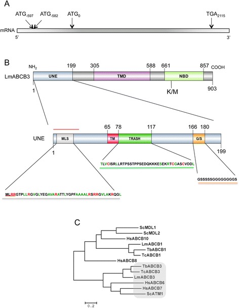
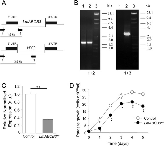
Results: LmABCB3 is an atypical ABC half-transporter that has a unique N-terminal extension not found in any other known ABC protein. This extension is required to target LmABCB3 to the mitochondrion and includes a potential metal-binding domain. We have shown that LmABCB3 interacts with porphyrins and is required for the mitochondrial synthesis of heme from a host precursor. We also present data supporting a role for LmABCB3 in the biogenesis of cytosolic ISC, essential cofactors for cell viability in all three kingdoms of life. LmABCB3 fully complemented the severe growth defect shown in yeast lacking ATM1, an orthologue of human ABCB7 involved in exporting from the mitochondria a gluthatione-containing compound required for the generation of cytosolic ISC. Indeed, docking analyzes performed with a LmABCB3 structural model using trypanothione, the main thiol in this parasite, as a ligand showed how both, LmABCB3 and yeast ATM1, contain a similar thiol-binding pocket. Additionally, we show solid evidence suggesting that LmABCB3 is an essential gene as dominant negative inhibition of LmABCB3 is lethal for the parasite. Moreover, the abrogation of only one allele of the gene did not impede promastigote growth in axenic culture but prevented the replication of intracellular amastigotes and the virulence of the parasites in a mouse model of cutaneous leishmaniasis.

Conclusions: Altogether our results present the previously undescribed LmABCB3 as an unusual mitochondrial ABC transporter essential for Leishmania survival through its role in the generation of heme and cytosolic ISC. Hence, LmABCB3 could represent a novel target to combat leishmaniasis.


Figures

Fig. 1
Fig. 1
LmABCB3 contains a unique N-terminal extension (UNE). a Schematic representation of LmABCB3 containing mRNA. In phase ATG codons found in 5‘ UTR and TGA Stop codon are indicated. b Schematic representation of LmABCB3 (up) showing the unique N-terminal extension (UNE), the Transmembrane Domain (TMD) and the Nucleotide Binding domain (NBD), indicating the K675M mutation (K/M) in the conserved Walker A motif that inactivates the protein. The schematic representation of the UNE region (down) highlights several motifs and sequences: i) a putative Mitochondrial Localization Signal (MLS) with conserved MLRR motif (underlined in red) and Arg (red) and hydrophobic Ala, Val and Leu residues (green); ii) a putative transmembrane segment (TM); iii) a TRASH domain with conserved Cys probably involved in metal co-ordination (red) and other residues (green) conserved in 70 % of TRASH domains [50]; iv) a Glycine/Serine repeat and iv) a putative signal peptide (red line). c Phylogenetic analysis of mitochondrial ABCB transporter sequences from Leishmania, mammals and yeast. Aligned protein sequences were subjected to phylogenetic analysis as described in Methods. The human representative of each mammalian subfamily was incorporated in the analysis to define each subfamily. Lm: L. major; Tb: T. brucei; Tc: T. cruzi; Hs: H. sapiens; Sc: S. cereviciae
Fig. 2
Fig. 2
The UNE region is required for the mitochondrial localization of LmABCB3. a Subcellular localization of LmABCB3. Representative picture of L. major promastigotes expressing LmABCB3-GFP or LmABCB3_∆UNE-GFP (GFP) were incubated at 28 °C with 50 nM of Mitotracker Red (Mit-Red) for 30 min at 4 °C. Nomarsky images are shown in the inset. Scale bar: 1 μm. The figure shows a representative parasite of a total population of parasites with a similar fluorescence pattern. b-c Quantitative colocalization analysis. Pearson’s coefficient (b) was calculated for each individual deconvolved image. Mander’s coefficients (c) were calculated to define the M1 and M2 as the proportion of pixels in one channel (M1 = green, GFP) that overlap with some signal in other channel (M2 = red, Mit-Red). Both coefficients were assessed using “JACoP”, a colocalization plugin available in Fiji software. Full: LmABCB3-GFP; ∆UNE: LmABCB3_∆UNE-GFP. ** p < 0.001
Fig. 3
Fig. 3
Generation and characterization of L. major promastigotes with one LmABCB3 allele deleted. a Schematic representation of the LmABCB3 locus and the hygromycin-resistance gene targeting construct used for gene replacement. The primers used (arrows 1–3) to verify the specific gene targeting and the expected sizes of the PCR-amplified products with the different pairs of primers are indicated. b PCR analysis of the LmABCB3 locus in control and single knock out (LmABCB3 +/−) mutant promastigotes. The specific gene-targeting PCR product (primers 1 and 3, 3.6 kb) confirmed that replacement with the hygromycin-resistance gene in one LmABCB3 allele in LmABCB3 +/−parasites (+/−, lanes 2). Primers 1 and 2 amplified the expected 1.6 kb product in Wt (+/+, lanes 1) and in LmABCB3 +/−parasites. Lanes 3 shows the DNA marker used. c LmABCB3 +/−promastigotes have reduced LmABCB3 expression. The expression level of LmABCB3 from control and LmABCB3 +/− L. major promastigotes was analyzed by qRT-PCR using mRNA isolated from each cell line as described in Methods. **p <0.001. d LmABCB3 +/− parasites grow as axenic promastigotes. Growth curve obtained after cultivation of control (white circles) and LmABCB3 +/−(black circles) promastigotes during the indicated time. The results represent the mean ± SEM of three independent experiments. *p < 0.05; **p < 0.001
Fig. 4
Fig. 4
LmABCB3 is required for the intracellular replication of amastigotes. a. Infection of macrophages by control and LmABCB3 ±/− parasites. Representative picture of the infection of THP-1 macrophages with control and LmABCB3 +/− stationary-phase L. major promastigotes, performed as described in Methods. At the indicated points, cells were fixed and DAPI (blue) stained. The macrophages nuclei (mn) and the kinetoplast and nuclei of intracellular amastigotes (arrowhead) are indicated. Scale bar: 5 um. b LmABCB3 +/− parasites have reduced ability to infect macrophages. THP-1 macrophages were infected with control and LmABCB3 +/−promastigotes as above described and the percentage of infected macrophages (n = 300 macrophages/group) was calculated. The results shown are the means ± SEM of three independent experiments performed in duplicated. **p <0.0002. c LmABCB3 +/−parasites are unable to replicate as intracellular amastigotes. THP-1 macrophages were infected with control and LmABCB3 +/− promastigotes as above described and the average number of intracellular amastigotes per infected macrophage was calculated at the indicated time points. The results shown are the means ± SEM. **p <0.0009
Fig. 5
Fig. 5
LmABCB3 is essential for L. major virulence. C57BL/6 male mice were infected with 1 × 106 stationary-phase L. major promastigotes of control or LmABCB3 +/− L. major in the left hind footpad. a Inflammation progression (difference between inoculated footpad and contralateral uninfected footpad) was recorded weekly. The values represent the means ± SEM of 7 mice. b Images show representative pictures of the footpad inflammation at 3 week post infection
Fig. 6
Fig. 6
LmABCB3 is required for mitochondrial heme biosynthesis. a LmABCB3 cell levels correlate with the amount of heme synthesized from exogenous PPIX. The mitochondrial synthesis of heme from its precursor PPIX was measured as described in Experimental procedures after incubation of control, LmABCB3 +/− and LmABCB3 overexpressing L. major promastigotes with 0.5 μM PPIX. The results represent the mean ± SEM of three independent experiments. * p<0.05, ** p<0.02, *** p<0.001. b LmABCB3 interacts with heme. Solubilized membrane proteins of L. major parasites overexpressing LmABCB3-GFP were subjected to a pull-down assay with hemin-agarose in the absence (0) or the presence of the indicated increasing concentration of free hemin (upper panel) or free PPIX (lower panel). LmABCB3-GFP was detected by inmmunoblotting with Anti-GFP antibody (1:5000)
Fig. 7
Fig. 7
LmABCB3 completely rescue the severe growth defect phenotype of yeast lacking ScATM1. a LmABCB3 allows the normal growth of ΔATM1 yeast in minimal medium. ΔATM1/ATM1 (control) and (ΔATM1/ATM1 + LmABCB3 (LmABCB3) cells were plated on minimal (SD) glucose media containing (+FOA) or not (−FOA) 1 mg/ml 5′-fluoroorotic acid (FOA) and incubated at 30 °C for three days. b LmABCB3 allows ΔATM1 yeast to use of non-fermentable carbon sources. ΔATM1, ΔATM1/ATM1 and ΔATM1/LmABCB3 cells were diluted into rich (YP) media containing 2 % (w/v) glucose, 3 % (w/v) galactose, 2 % (v/v) glycerol, 2 % (w/v) lactate, or 2 % (v/v) ethanol to an A600 0.05. After 24 (left) or 48 (right) hours of growth at 30 8C, the A600 was measured. Growth is expressed relative to ΔATM1/ATM1 growth in HC glucose
Fig. 8
Fig. 8
LmABCB3 homology model shares a thiol binding region with ScATM1 (a). Structural homology among LmABCB3 and ScATM1. A model for the 3D-structure of L. major ABCB3_∆UNE (aa 283–875) was built using Phyre2 molecular modelling server based on LmABCB3_∆UNE complete protein sequence, its predicted secondary structure and protein structures present in the PDB (Protein Data Bank) database. The structure of yeast ATM1 (pdb code: 1 MHY) was chosen by the server as the less divergent to the constructed LmABCB3 model with a 40 % sequence similarity. 3D-structural superimposition of yeast ATM1 structure (represented as yellow ribbons) and L. major ABCB3_∆UNE model (showed in red ribbons) was done using the DALI server (Z score: 26.5; RMSD:3.5 Å). b LmABCB3 conserves a positively charged pocket similar to the glutathione binding cavity present in ScATM1. Molecular surface representation colored by electrostatic charges (blue, positive; red, negative) distribution of yeast ATM1 and L. major ABCB3_∆UNEmodel. A positively charged pocket which binds a GSH molecule in ScATM1 is highlighted. An equivalent positive region is also observed in the proposed LmABCB3 model. c Trypanothione binding region in LmABCB3 dimer. Left. A model of LmABCB3_∆UNE dimer was built with chains A and B colored in red and green respectively. The residues conforming the T(SH)2 binding site of both monomers are highlighted in blue dots representation. Right. Molecular docking of reduced trypanothione (two molecules colored yelow) into the molecular surface of L. major ABCB3 dimer model (chains A and B colored red and green respectively) using Autodock4.0 program. d Close view of potential trypanothione binding site. The T(SH)2 molecule (colored green) appears surrounded by LmABCB3 conserved residues (yellow) in the conserved cavity of LmABCB3 model (monomer represented in red sticks)

References

    1. Pace D. Leishmaniasis. J Infect. 2014;69:S10–18. doi: 10.1016/j.jinf.2014.07.016. - DOI - PubMed
    1. McCall LI, McKerrow JH. Determinants of disease phenotype in trypanosomatid parasites. Trends Parasitol. 2014;30(7):342–349. doi: 10.1016/j.pt.2014.05.001. - DOI - PubMed
    1. DNDi: http://www.dndi.org/diseases-projects/leishmaniasis/.
    1. Trager W. Nutrition and biosynthetic capabilities of flagellates. Problems of in vitro cultivation and differentiation. In: CIBAFoundationSymposium, editor. Trypanosomiasis and Leishmaniasis with special reference to Chagas’ disease. North Holland, Amsterdam: Elsevier-Excerpta; 1974. pp. 225–245.
    1. Opperdoes FR, Coombs GH. Metabolism of Leishmania: proven and predicted. Trends Parasitol. 2007;23(4):149–158. doi: 10.1016/j.pt.2007.02.004. - DOI - PubMed

Publication types

LinkOut - more resources