

doi: 10.1038/srep18734.
Transgenic FingRs for Live Mapping of Synaptic Dynamics in Genetically-Defined Neurons
Affiliations
- PMID: 26728131
- PMCID: PMC4700522
- DOI: 10.1038/srep18734
Item in Clipboard
Transgenic FingRs for Live Mapping of Synaptic Dynamics in Genetically-Defined Neurons
Sci Rep.
.
Abstract
Tools for genetically-determined visualization of synaptic circuits and interactions are necessary to build connectomics of the vertebrate brain and to screen synaptic properties in neurological disease models. Here we develop a transgenic FingR (fibronectin intrabodies generated by mRNA display) technology for monitoring synapses in live zebrafish. We demonstrate FingR labeling of defined excitatory and inhibitory synapses, and show FingR applicability for dissecting synapse dynamics in normal and disease states. Using our system we show that chronic hypoxia, associated with neurological defects in preterm birth, affects dopaminergic neuron synapse number depending on the developmental timing of hypoxia.
Figures
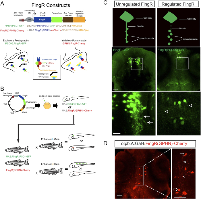
(A) Schematic diagram of FingR constructs used to generate plasmids and transgenic lines. FingR(PSD95)-GFP binds endogenous PSD-95 protein at the post-synaptic density; FingR(GPHN)-Cherry binds endogenous GPHN. (B) Use of inducible Gal4/UAS system with FingR(GPHN) or FingR(PSD95) in different Gal4 lines for differential synapse labeling (drawn by JHS). (C) Contrasting examples of unregulated (FingR-GFP) and regulated (FingR(PSD95)-GFP) FingR plasmids injected into Tg(otpb.A.Gal4) embryos. GFP signal from the unregulated FingR is distributed throughout the neuron and neurites (arrows) and individual synaptic puncta are not visualizable. In contrast, distinct puncta are seen (open arrowheads) using regulated FingR expression. Confocal images, scale bar 50 μm, 10 μm in inset panels; confocal z-stacks, ventral views, rostral to the top. D) Demonstration of puncta labeling with regulated FingR for GPHN. Confocal z-stacks, ventral view, rostral to top, of Tg(otpb.A:Gal4) embryo injected with FingR(GPHN)-Cherry. Arrows, neuron soma; arrowheads, puncta. Scale bar 50 μm, 10 μm in inset panel.
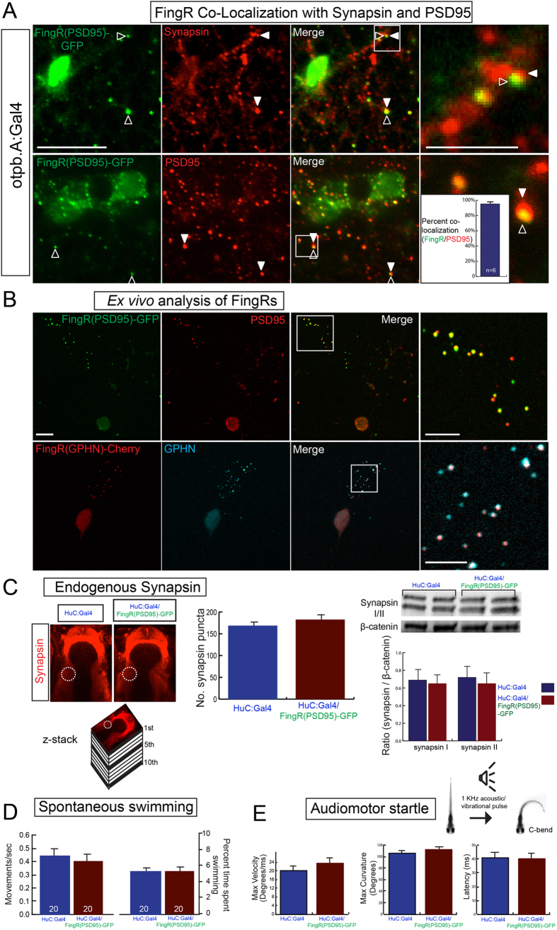
(A) FingR(PSD95)-GFP signal is adjacent to Synapsin immunohistochemistry (top panels) and overlaps PSD-95 immunohistochemistry (bottom panels). Quantification of FingR(PSD95)-GFP signal and endogenous PSD-95 signal demonstrates 95% overlap of PSD-95 immunohistochemistry with GFP signal from FingR (bar graph: median 95% +/− 1%, SEM; n = 6 larvae; p 0.01). Confocal images of sections from immunostained Tg(otpb.A:Gal4); Tg(FingR(PSD95)-GFP) larvae, scale bar 10 μm, 5 μm in inset panels. (B) Ex vivo sparse zebrafish primary neuron cell culture demonstrates co-localization of FingRs and endogenous synaptic proteins. Top row, dissociated neurons from Tg(otpb.A:Gal4); Tg(FingR(PSD95)-GFP) embryo, immunohistochemistry for anti-GFP and anti-PSD95. Bottom row from Tg(HuC:Gal4); Tg(FingR(GPHN)-Cherry) embryo, immunohistochemistry for anti-mCherry and anti-GPHN. Confocal images of slides, scale bar 10 μm, 5 μm in inset. (C–E) Pan-neuronal expression in Tg(FingR(PSD95)-GFP); Tg(HuC:Gal4) larvae. (C) FingR(PSD95)-GFP expression does not affect Synapsin protein expression. Data is double-transgenic larvae compared to Tg(HuC:Gal4) only. Images are confocal z-stacks, dorsal views, rostral to top; dotted circle indicates area of Synapsin puncta quantification for bar graphs. Western blots are for Synapsin in whole larvae, standardized to β-catenin; quantified bar graphs to right. (D) FingR(PSD95)-GFP expression does not affect spontaneous swimming behavior (percent time swimming and number of movements), n = 20 embryos each test; three separate experiments; SEM. (E) FingR(PSD95)-GFP expression does not affect audiomotor startle response, n = 27 (control) and 30 (FingR) embryos each test; three separate experiments, SEM (velocity, body curvature, or latency).
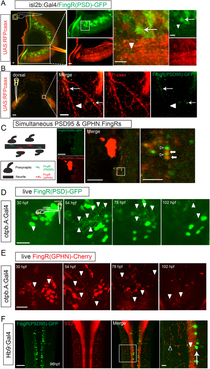
(A) Lateral view of eye from transgenic zebrafish expressing FingR(PSD95)-GFP and RFP-caax in retinal ganglion neurons (transgenic Tg(isl2b:Gal4); Tg(UAS:RFP-caax); Tg(FingR(PSD95)-GFP)). GFP expression is seen in puncta (arrowheads) and neuron somas (arrow); RFP labels neurons, dendrites, and their axons as they project towards optic chiasm (Supplemental Movie 1). Confocal z-stacks at 72 hpf, immunohistochemistry for GFP and RFP, scale bar 10 μm. (B) Dorsal view of tectum in Tg(isl2b:Gal4); Tg(UAS:RFP-caax); Tg(FingR(PSD95)-GFP) embryo. Arrow shows FingR(PSD95)-GFP puncta along RGC axon, but not along entire length of axon (arrowhead). Confocal z-stack at 72 hpf, immunohistochemistry for GFP and RFP; scale bar 50 μm, 5 μm in inset panel. (C) Larvae co-expressing FingR(PDS95)-GFP (open arrowheads) and FingR(GPHN)-Cherry (arrows) demonstrates presence of neighboring excitatory and inhibitory puncta. Confocal z-stacks; scale bar 10 μm, 2.5 μm in inset panel. (D,E) FingRs can be used for in vivo visualization and monitoring of synapses. (D) Time-series of dopaminergic neurons and synapses in Tg(otpb.A:Gal4); Tg(FingR(PSD95)-GFP) animal, demonstrating changes in synapse number and expression with development. Scale bar 20 μm. (E) Time-series of dopaminergic neurons and synapses development in Tg(otpb.A:Gal4); Tg(FingR(GPHN)-Cherry) animal, demonstrating changes in synapse number and expression with development. Scale bar 20 μm. (F) Dorsal view in trunk at 96 hpf showing that FingR(PSD95)-GFP is expressed in the spinal cord (arrowhead), but not in axon projections labeled with SV2 (open arrowhead) in the neuromuscular synapses when driven in motor neurons (arrow) by Hb9:Gal4. Confocal z-stacks at 96 hpf, dorsal views, immunohistochemistry for GFP and SV2; scale bar 50 μm, 10 μm in inset panel.
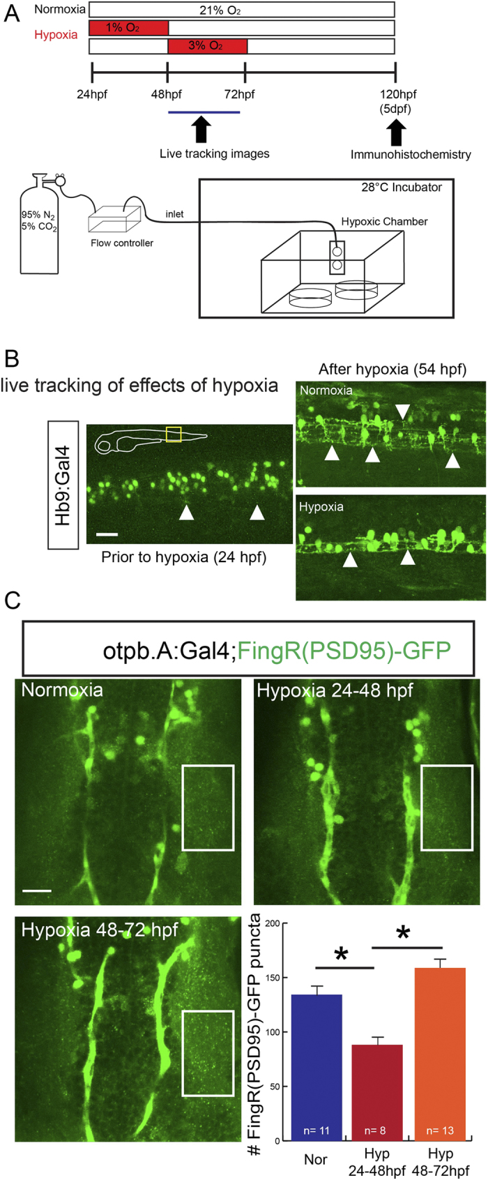
(A) Schematic illustration of hypoxia exposure and imaging. (B) Demonstration of live imaging of effects of hypoxic injury on motor neuron synaptic puncta visualized by FingR(PSD)-GFP in trunk of Tg(Hb9:Gal4); Tg(FingR(PSD95)-GFP) animals. Following hypoxia there is a decrease in the number of puncta labeled by FingR(PSD95)-GFP. Confocal images, rostral to left, scale bar 10 μm. (C) Confocal images and quantification in the dopaminergic neurons in the diencephalon of Tg(otpb.A:Gal4); Tg(FingR(PSD95)-GFP) animals. Hypoxia from 24–48 hpf decreased number of puncta but later hypoxia exposure did not significantly change number; (p = 0.002; one-way ANOVA; SEM shown). Confocal z-stacks, rostral to top, scale bar 10 μm.
Similar articles
-
A Viral Toolbox of Genetically Encoded Fluorescent Synaptic Tags.iScience. 2020 Jul 24;23(7):101330. doi: 10.1016/j.isci.2020.101330. Epub 2020 Jun 30. iScience. 2020. PMID: 32674057 Free PMC article.
-
Directional Trans-Synaptic Labeling of Specific Neuronal Connections in Live Animals.Genetics. 2015 Jul;200(3):697-705. doi: 10.1534/genetics.115.177006. Epub 2015 Apr 27. Genetics. 2015. PMID: 25917682 Free PMC article.
-
A transgenic zebrafish model for in vivo long-term imaging of retinotectal synaptogenesis.Sci Rep. 2018 Sep 19;8(1):14077. doi: 10.1038/s41598-018-32409-y. Sci Rep. 2018. PMID: 30232367 Free PMC article.
-
Transgenic technology for visualization and manipulation of the neural circuits controlling behavior in zebrafish.Dev Growth Differ. 2008 Jun;50 Suppl 1:S167-75. doi: 10.1111/j.1440-169X.2008.01003.x. Epub 2008 Apr 22. Dev Growth Differ. 2008. PMID: 18430169 Review.
-
Fluorescence imaging of synapse formation and remodeling.Microscopy (Oxf). 2013 Feb;62(1):51-62. doi: 10.1093/jmicro/dfs083. Epub 2012 Dec 14. Microscopy (Oxf). 2013. PMID: 23243097 Review.
Cited by
-
Hypoxia and connectivity in the developing vertebrate nervous system.Dis Model Mech. 2018 Dec 12;11(12):dmm037127. doi: 10.1242/dmm.037127. Dis Model Mech. 2018. PMID: 30541748 Free PMC article. Review.
-
Towards a Comprehensive Optical Connectome at Single Synapse Resolution via Expansion Microscopy.Front Synaptic Neurosci. 2022 Jan 18;13:754814. doi: 10.3389/fnsyn.2021.754814. eCollection 2021. Front Synaptic Neurosci. 2022. PMID: 35115916 Free PMC article. Review.
-
Dopaminergic Co-Regulation of Locomotor Development and Motor Neuron Synaptogenesis is Uncoupled by Hypoxia in Zebrafish.eNeuro. 2020 Feb 27;7(1):ENEURO.0355-19.2020. doi: 10.1523/ENEURO.0355-19.2020. Print 2020 Jan/Feb. eNeuro. 2020. PMID: 32001551 Free PMC article.
-
Hypoplasia of dopaminergic neurons by hypoxia-induced neurotoxicity is associated with disrupted swimming development of larval zebrafish.Front Cell Neurosci. 2022 Sep 23;16:963037. doi: 10.3389/fncel.2022.963037. eCollection 2022. Front Cell Neurosci. 2022. PMID: 36212692 Free PMC article.
-
Controlling ion channel function with renewable recombinant antibodies.J Physiol. 2022 May;600(9):2023-2036. doi: 10.1113/JP282403. Epub 2022 Mar 17. J Physiol. 2022. PMID: 35238051 Free PMC article. Review.
References
Publication types
MeSH terms
Substances
Grants and funding
LinkOut - more resources
Full Text Sources
Other Literature Sources
Molecular Biology Databases
Research Materials
